

Alita Loveless, MD
- Instructor
- Department of Obstetrics and Gynecology
- Texas Tech University Health Sciences Center
- Lubbock, Texas
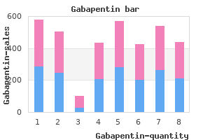
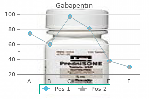
Levoxine symptoms qt prolongation buy gabapentin master card, Levoxyl medications affected by grapefruit cheapest gabapentin, Synthroid) that is used as a thy roid hormone replacement drug to treat an under ligament treatment 1st line order gabapentin with visa, posterior cruciate See posterior active thyroid gland (hypothyroidism) treatment yellow fever cheap gabapentin 100mg on line. The abnormal areas on the skin in lichen lightheadedness A feeling that one is about to planus are typically flat-topped (hence the term faint section 8 medications generic gabapentin 800 mg without prescription. Lightheadedness is medically distinct from planus) treatment 0 rapid linear progression buy gabapentin 100 mg visa, itchy, and frequently have a polygonal or dizziness, unsteadiness, and vertigo. Lichen planus lights, flashing A sensation that is created when on the scalp may lead to hair loss. The causes the clear, jelly-like substance that fills the middle of of lichen planus are unknown. However, it can be the eye (vitreous humor) shrinks and tugs on the triggered by the use of certain drugs, such as thi retina. These flashes of light can appear off and on azide diuretics, phenothiazines, and antimalarials. Some of them are present at birth (congenital), and others are limb An arm or a leg. In lipodystrophy syndrome, the face, together as a package because of their location near arms, and legs become thin due to loss of subcuta one another on the same chromosome. See also cephalothoracic lipody lip One of the two fleshy folds that surround the strophy; protease inhibitor. The upper lip is separated from the nose by the philtrum, the area that lies lipoma A benign tumor of adipocytes (fat cells). Small blind pits lipomatosis, familial benign cervical See are sometimes seen at the corners of the mouth; cephalothoracic lipodystrophy. The lips may be abnor lipoprotein A molecule that is a combination of mally thin or thick. Lipids do not travel in the blood alcohol syndrome typically have a thin upper lip and by themselves, but they are carried through the flat philtrum. Most frequently seen in older adults (age 40 and above), liposarcomas are the most common of lipid A fat. Lipids are easily stored in the body liposuction the surgical suctioning of fat and serve as fuel. Among the well-known lipids are deposits from specific parts of the body, the most cholesterol, triglycerides, fatty acids, and steroids common being the abdomen, buttocks, hips, thighs (such as cortisone). A phospholipids are all compound lipids (lipids in hollow instrument called a cannula is inserted combination with other types of chemicals). A high-pressure vacuum is then applied to the cannula to suck out lipid profile A pattern of lipids in the blood. Lipid storage diseases result in the abnormal includes the local anesthetic lidocaine to numb accumulation of lipids in various organs. Examples the area and the vessel-constrictor epinephrine include Gaucher disease, Fabry disease, Niemann (adrenaline) to help minimize bleeding. This technique has an advantage in areas of scar tissue, such as the male breast, the back, and lithotomy Surgical removal of a stone. Its disadvantages include the need for lithotripsy A procedure that uses shock waves to longer incisions in the skin, a potential for skin or break a stone in the kidney, urinary tract, or gall internal burns, greater cost, and a longer time bladder. Anesthesia may be necessary to control the pain, Listeriosis A disease that is caused by eating depending on the size and density of the stone and food contaminated with the bacterium Listeria on the energy of the shock wave needed to break it monocytogenes. The urologist may opt to place a catheter (stent) health problem in North America. The disease in the ureter from below to facilitate passage of the affects primarily pregnant women, newborns, and shattered fragments. The Listeriosis can prevent the infection by avoiding cer procedure is done under anesthesia, using real tain high-risk foods and by handling food properly. Raw food from animal sources (such as beef, pork, or poultry) should be thoroughly cooked and lithotriptor A machine that is used to shatter uncooked meats should be kept separate from veg kidney stones and gallstones by physical or other etables, cooked foods, and ready-to-eat foods. A liter is a little more been reported in association with autoimmune dis than 1 quart (1. Treatment of lithium toxicity store, and process fats, including fatty acids (used involves immediately reducing or discontinuing for energy) and cholesterol; to metabolize and store carbohydrates (used as the source for the sugar in lithium use under medical supervision. The most common rea fissures, connective tissue, or other natural bound son for liver transplantation in children is biliary aries. The most common reason for liver transplantation in adults is lobectomy An operation to remove an entire cirrhosis (a disease in which healthy liver cells are lobe of the lung. There is no effective treatment for end-stage liver disease other lobular carcinoma of the breast, infiltrating than transplantation. At first the specifies what types of medical treatment are lochia is primarily blood, followed by a more desired. A living will can be very specific or very mucousy fluid that contains dried blood, and finally general. The irregular heartbeats are typically brought lower segment cesarean section See cae on by stress or vigorous activity. The lumbar vertebrae and their longitudinal section A section that is cut along disks are situated below the thoracic vertebrae and the long axis of a structure. Longitudinal section is above the sacral vertebrae in the spinal column and the opposite of cross-section. For example, a longitudinal study of lumbar puncture A procedure in which cere children with Down syndrome might involve the brospinal fluid is removed from the spinal canal for study of 100 children with this condition from birth diagnostic testing or treatment. Longitudinal study is the opposite patient usually lies sideways for the procedure, of cross-sectional (synchronic) study. As inflammation continues, scar tissue may done for therapeutic purposes, as a way of adminis form, including keloid scarring in patients prone to tering antibiotics, cancer drugs, or anesthetic agents keloid formation. Lupus is more common in patients with conditions such as normal-pressure women than in men, and although it occurs in all hydrocephalus or benign intracranial hypertension. These complications vation of symptoms, and through testing of the are uncommon, with the exception of headache, blood for signs of autoimmune activity. Headaches ment is essential to prevent progression of the dis are less likely to occur if the patient remains lying ease. A rheumatologist can provide treatment for flat for 1 to 3 hours after the procedure. Also known lupus, and this treatment has two objectives: treat as spinal tap, spinal puncture, thecal puncture, and ing the difficult symptoms of the disease and treat rachiocentesis. It may include use of steroids and other anti-inflammatory lumbar vertebrae the five vertebrae situated agents, antidepressants and/or mood stabilizers, between the thoracic vertebrae and the sacral verte intravenous immunoglobulin, and, in cases in brae in the spinal column. The lumbar vertebrae are which lupus involves the internal organs, represented by the symbols L1 through L5. In com lupus, discoid A chronic inflammatory condi mon use, lumpectomy refers especially to removal tion that is limited to the skin and is caused by an of a lump from the breast. Skin symp toms associated with discoid lupus include patchy lung One of a pair of three-lobed breathing redness with areas of hyper and hypopigmentation organs located within the right and left sides of the that can cause scarring; and photosensitivity, or skin chest. The lungs remove carbon dioxide from the rash in reaction to exposure to sunlight. Treatment the left and right lungs by means of the left and right is directed toward decreasing inflammation and/or bronchi. Treatment methods smaller bronchioles, which end in many alveolar include avoidance of sun exposure and use of anti sacs. In the tiny alveoli within these sacs, oxygen is malarial medications (hydroxychloroquine and oth exchanged for carbon dioxide in blood delivered ers), local cortisone injections, Dapsone, and back to the heart by the pulmonary veins. See also lupus; function is controlled by several muscles, including lupus erythematosis, systemic. Lung transplant is sometimes done in tan exposure; ulceration of the mucus lining of the dem with heart transplant. Patients with lupus have in carditis/pleuritis), usually associated with chest pain their blood unusual antibodies that are targeted with breathing; abnormal amounts of protein or cel against their own body tissues. Lupus can cause dis lular elements in the urine, caused by kidney abnor ease of the skin, heart, lungs, kidneys, joints, and malities; brain irritation manifested by seizures, nervous system. The first symptom is a red (or severe mood swings, and/or psychosis; low counts of dark), scaly rash on the nose and cheeks, often white or red blood cells, or platelets; abnormal. Psychiatric symptoms closely resem Within hours to weeks of the tick bite, an expanding ble those of a bipolar disorder, which sometimes ring of unraised redness develops, with an outer leads to misdiagnosis. The red unknown, but heredity, infectious disease, ultraviolet ness of the skin is often accompanied by generalized light, and drugs may all play a role. Treatment is fatigue, muscle and joint stiffness, swollen glands, directed toward decreasing inflammation and mod and headache. Early treatment with antibiotics is the erating the level of autoimmune activity, and it can best strategy for preventing major problems due to range from administration of anti-inflammatory Lyme disease. Medication can help treat spe Lyme disease only became apparent in 1975, when cific symptoms as well, including reducing skin rash, mothers of a group of children who lived near each irritation, and scarring; reducing joint inflammation; other in Lyme, Connecticut, made researchers and treating psychiatric symptoms. The sen hormone that controls the production of luteinizing tinel node for a given tumor is found by injecting a hormone in men and women. A partial that can be tracked visually or a radioactive colloid dislocation is a subluxation. Biopsy of the sentinel lymph node can reveal whether cancer has Lyme disease An inflammatory disease that is spread through the lymphatic system. If the sentinel caused by the bacterium Borrelia burgdorferi, node contains tumor cells, removal of more nodes which is transmitted to humans by the deer tick. In addition, these patients can have pain around the anal area (perianal), and occasionally lymphangioma An abnormal structure that con drainage from the perianal area or the glands in the sists of a collection of blood vessels and lymph ves groin. Depending on its nature, a lymphangioma may grow lymphoid Referring to lymphocytes, a type of slowly or quickly. Lymphangiomas can cause prob white blood cell, or to tissue in which lymphocytes lems because of their location. Lymphoid tissue is full of lymphocytes, phangioma around the larynx might cause a such as a lymph node. Lymphoid tissue is present throughout the body ultimately drains back into the bloodstream. Treatment options include chemo and may occur in the arm or leg after lymph vessels or radiation therapy. Treatment is like that of other lym Lymphocytes are integrally involved in many phomas but must take into account the fact that the immune responses. For example, lymphocytic inflam tissue that is characterized by unusually large cells mation in the skin is skin that is infiltrated with when viewed microscopically. Since it is a tumor con lymphocytopenia, ranging from medication toxicity to sisting of early lymphocyte precursors (lym a variety of diseases. Lymphoblastc lymphoma is a term that has Lymphocytosis may be a marker that infection or been used in the past to refer to the presence of the disease is present. Treatment may include chemotherapy, radi eases of the lymphoid cells and of cells from the ation, surgery, medications, and bone marrow reticuloendothelial system that usually occur in transplant. Diagnosis is sis (the destruction of red blood cells with the made via biopsy of a swollen lymph node, although release of hemoglobin). Treatment may include chemother lytic Suffix having to do with lysis, as in apy, radiation, bone marrow transplantation, stem hemolytic anemia (anemia due to the destruction of cell transplantation, use of medication, and the use red blood cells). For example, macrocytic ane mia is characterized by abnormally large red blood cells. Macrogenitosomia is associated with hor monal disorders that may also create changes in the internal sex organs. The urine of patients with multiple myeloma, a form of large protein is an antibody called macroglobulin or cancer that arises in plasma cells. Macroglossia is sometimes said to be associated with Macewen operation A surgical operation to Down syndrome, but in that disorder the tongue is repair inguinal hernia that was designed by Scottish actually large only in relationship to a smaller-than surgeon Sir William Macewen. To brovascular disease (macroangiopathy in the determine if the testes are too large, a device called brain), and peripheral vascular disease (macroan an orchidometer is used that permits a testis to giopathy that affects, for example, vessels in the be compared to a series of plastic ovals (like minia legs). The opposite macrobiotic Referring to the macrobiota, a of macroorchidism is microorchidism. Macrophages are key play macrobiotic diet A diet that incorporates ers in the immune response to foreign invaders of Ayurvedic principles of food combining, is based the body, such as infectious microorganisms. They mainly on brown rice and vegetables, and claims to are normally found in the liver, spleen, and connec lengthen life. For exam ple, a macroscopic tumor is big enough to see with macrocephaly An abnormally large head. Macrocephaly can be a normal variant or be a sign macrosomia An overly large body.

A symptoms 24 hour flu cheap gabapentin online visa, Cross-section of aortic media with marked elastin fragmentation and formation of areas devoid of elastin that resemble cystic spaces medications vascular dementia buy cheap gabapentin on-line, from a patient with Marfan syndrome symptoms torn meniscus order gabapentin discount. B medicine 377 cheap gabapentin online, Normal media for comparison treatment 4 ringworm discount gabapentin 800mg without a prescription, showing the regular layered pattern of elastic tissue treatment yeast infection men purchase 400mg gabapentin free shipping. The serious complications predominantly occur in the region from the aortic valve through the arch. The main immunologic mechanisms that initiate noninfectious vasculitis are: (1) immune complex deposition, (2) antineutrophil cytoplasmic antibodies, and (3) anti-endothelial cell antibodies. Immune reactants and complement can be detected in the serum or vessels of patients with vasculitis. Some, such as penicillin, conjugate serum proteins; others, like streptokinase, are themselves foreign proteins. The manifestations vary and range from small-vessel hypersensitivity and leukocytoclastic vasculitis to polyarteritis nodosa, Wegener granulomatosis, and Churg-Strauss syndrome (see later for descriptions of these entities), and from mild and self-limiting to severe and even fatal. Whether immune complexes deposit in vessel walls from the circulation, or are formed in situ, or both, is not known (see Chapter 6). However, many small vessel vasculitides show a paucity of vascular immune deposits and therefore other mechanisms have been sought for these so-called pauci-immune vasculitides. The description of these autoantibodies is based on the immunofluorescent patterns of staining of ethanol-fixed neutrophils. The systemic vasculitides are classified on the basis of the size and anatomic site of the involved blood vessels (. There is considerable clinical and pathologic overlap among these disorders summarized in Table 11-5 (Table Not Available) and discussed below. Therefore, visual loss caused by giant cell arteritis is a medical emergency that requires prompt recognition Figure 11-23 Diagrammatic representation of the sites of the vasculature involved by the major forms of vasculitis. The widths of the trapezoids indicate the frequencies of involvement of various portions. A, H&E stain of section of temporal artery showing giant cells at the degenerated internal elastic membrane in active arteritis (arrow). C, Examination of the temporal artery of a patient with giant-cell arteritis shows a thickened, nodular, and tender segment of a vessel on the surface of head (arrow). A, Aortic arch angiogram showing narrowing of brachiocephalic, carotid, and subclavian arteries (arrows). B, Gross photograph of two cross-sections of the right carotid artery taken at autopsy of the patient shown in A, demonstrating marked intimal thickening with minimal residual lumen. C, Histologic view of active Takayasu aortitis, illustrating destruction of the arterial media by mononuclear inflammation with giant cells. Figure 11-26 Representative forms of systemic medium-sized to small vessel vasculitis. In polyarteritis nodosa (A), there is segmental fibrinoid necrosis and thrombotic occlusion of the lumen of this small artery. In leukocytoclastic vasculitis (B), shown here from a skin biopsy, there is fragmentation of neutrophils in and around blood vessel walls. In Wegener granulomatosis (C), there is inflammation (vasculitis) of a small artery along with adjacent granulomatous inflammation, in which epithelioid cells and giant cells (arrows) are seen. D, Gross photo from the lung of a patient with fatal Wegener granulomatosis, demonstrating large nodular lesions. In a typical case of Buerger disease (E), the lumen is occluded by a thrombus containing two abscesses (arrow). A, Sharply demarcated pallor of the distal fingers resulting from the closure of digital arteries. Because these lesions constitute abnormalities of unregulated vascular proliferation, the possibility of controlling such growth by agents that inhibit blood vessel formation (anti-angiogenic factors) is particularly exciting. The majority are superficial lesions, often of the head or neck, but they may occur internally, with nearly one third in the liver. Hemangiomas constitute 7% of all benign tumors in infancy and childhood (Chapter 10). Nevertheless, many of the capillary lesions regress spontaneously at or before puberty. Capillary hemangiomas, the largest single type of vascular tumor, are most common in the skin, subcutaneous tissues, and mucous membranes of the oral cavities and lips, but they may also occur in the liver, spleen, and kidneys. The "strawberry type" of capillary hemangioma (juvenile hemangioma) of the skin of newborns is extremely common (1 in 200 births), may be multiple, grows rapidly in the first few months, begins to fade when the 546 Figure 11-30 Hemangiomas. B, Histologic appearance with acute neutrophilic inflammation and vascular (capillary) proliferation. Inset, demonstration by modified silver (Warthin-Starry) stain of clusters of tangled bacilli (black). A, Gross photograph, illustrating coalescent red-purple macules and plaques of the skin. B, Histology of nodular form, demonstrating sheets of plump, proliferating spindle cells. B, Photomicrograph of moderately well-differentiated angiosarcoma with dense clumps of irregular, moderate anaplastic cells and distinct vascular lumens. A, Coronary artery with recent balloon angioplasty, in a low-power photomicrograph showing the split encompassing the intima and media (arrow) and partial circumferential dissection. B, Gross photograph of restenosis following balloon angioplasty, demonstrating residual atherosclerotic plaque (left arrow) and a new, glistening proliferative lesion (right arrow). C, Coronary arterial stent implanted long term, demonstrating thickened neointima separating the stent wires (black spot shown by arrow) from the lumen (asterisk). Pathology of cardiovascular interventions, including endovascular therapies, revascularization, vascular replacement, cardiac assist/replacement, arrhythmia control, and repaired congenital heart disease. B, Photomicrograph demonstrating Gore-Tex graft (arrow) with prominent intimal proliferation and very small residual lumen (asterisk). Shin D, et al: Expression of ephrin-B2 identifies a stable genetic difference between arterial and venous vascular smooth muscle as well as endothelial cells, and marks subsets of microvessels at sites of adult neovascularization. Garcia-Cardena G, et al: Biomechanical activation of vascular endothelium as a determinant of its functional phenotype. Folkman J, et al: Angiogenesis research: guidelines for translation to clinical application. Angelini P, et al: Coronary anomalies: incidence, pathophysiology, and clinical relevance. Geng Y-J, Libby P: Progression of atheroma: a struggle between death and procreation. Corti R, et al: Vasopeptidase inhibitors: a new therapeutic concept in cardiovascular disease Knox J, et al: Evidence for altered balance between matrix metalloproteinases and their inhibitors in human aortic diseases. Salvarani C, et al: Polymyalgia rheumatica and giant-cell arteritis N Engl J Med 347:261, 2002. As might be anticipated, cardiac dysfunction can be associated with devastating physiologic consequences. Heart disease is the predominant cause of disability and death in industrialized nations. In the United States, it accounts for about 40% of all postnatal deaths, totaling about 750, 000 individuals annually and nearly twice the number of deaths caused by all forms of cancer combined. The yearly economic burden of ischemic heart disease, the most prevalent subgroup, is estimated to be in excess of $100 billion. The major categories of cardiac diseases considered in this chapter include congenital heart abnormalities, ischemic heart disease, heart disease caused by systemic hypertension, heart disease caused by pulmonary diseases (cor pulmonale), diseases of the cardiac valves, and primary myocardial diseases. A few comments about pericardial diseases and cardiac neoplasms as well as cardiac transplantation are also offered. Before considering details of specific conditions, we will review salient features of normal anatomy and function as well as the principles of cardiac hypertrophy and failure, the common end points of many different types of heart disease. Normal the normal heart weight varies with body height and weight; it averages approximately 250 to 300 g in females and 300 to 350 g in males. As will be seen, increases in cardiac size and weight accompany many forms of heart disease. Greater heart weight or ventricular thickness indicates hypertrophy, and an enlarged chamber size implies dilation. An increase in cardiac weight or size (owing to hypertrophy and/or dilation) is termed cardiomegaly. They are arranged largely in a circumferential and Figure 12-1 Myocardium (cardiac muscle). A the histology of myocardium is shown, emphasizing the centrally-placed nuclei of the cardiac myocytes (arrowhead), intercalated discs (representing specialized end-to-end junctions of adjoining cells; highlighted by a double arrow) and the sarcomeric structure visible as cross-striations within myocytes. Triphenyltetrazolium staining of irreversible injury following coronary artery occlusion in rats. Superficial endothelial cells (arrow) and diffusely distributed deep interstitial cells are noted. The strength of the valve is predominantly derived from the fibrosa, with its dense collagen (yellow). This section highlights the dense, laminated elastic tissue in the ventricularis (double arrow). A reduction in the size of the left ventricular cavity, particularly in the base-to-apex dimension, is associated with increasing age and accentuated by systemic hypertension. Accompanied by a rightward shift and tortuosity of a dilated ascending aorta, this chamber alteration causes the basal ventricular septum to bend leftward, bulging into the left ventricular outflow tract (termed sigmoid septum). Such reduction in the size of the left ventricular cavity can simulate the obstruction to blood leaving the left ventricle that often occurs with hypertrophic cardiomyopathy, discussed later in this chapter. Several changes of the valves are noted with aging, including calcification of the mitral annulus and aortic valve, the latter frequently leading to aortic stenosis. In addition, the valves can develop fibrous thickening, and the mitral leaflets tend to buckle back toward the left atrium during ventricular systole, simulating a prolapsing (myxomatous) mitral valve (see later). Moreover, many older persons develop small filiform processes (Lambl excrescences) on the closure lines of aortic and mitral valves, probably arising from the organization of small thrombi on the valve contact margins. Compared with younger myocardium, "elderly" myocardium also has fewer myocytes, increased collagenized connective tissue and, in some individuals, deposition of amyloid. In the muscle cells, lipofuscin deposits (Chapter 1), and basophilic degeneration, an accumulation within cardiac myocytes of a gray-blue byproduct of glycogen metabolism, may be present. Extensive lipofuscin deposition in a small, atrophied heart is called brown atrophy; this change often accompanies cachectic weight loss, as seen in terminal cancer. Although the morphologic changes described are common in elderly patients at necropsy, and they may mimic disease, in only a minority are they associated with clinical cardiac dysfunction. In the most common circumstance, the cardiac muscle contracts weakly or inadequately, and the chambers cannot empty properly. In some conditions, however, the muscle cannot relax sufficiently to permit ventricular filling. Most cardiovascular disease arises from the interaction of environmental factors and genetic susceptibility. The contemporary view holds that most clinical cardiovascular diseases result from a complex interplay of genetics and environmental factors that disrupt networks controlling morphogenesis, myocyte survival, biomechanical stress responses, contractility, and [8] electrical conduction. For example, there is growing recognition that pathogenesis of congenital heart defects, in many cases, involves an underlying genetic abnormality whose expression is strongly modified by external (environmental or maternal) factors. Moreover, since a diverse group of cytoskeletal protein mutations have been linked with cardiac muscle cell dysfunction in the cardiomyopathies, perhaps subtle mutations or polymorphisms in these genes could confer an increased risk or more rapid onset of heart failure in response to acquired cardiac injury. In these and other examples, the clinical expression of cardiac disease represents the end result of multiple internal and external cues for growth, death, and survival [9] of cardiac myocytes. These factors and pathways are shared with other normal tissues and pathological processes. Although usually caused by a slowly developing intrinsic deficit in myocardial contraction, a similar clinical syndrome is present in some patients with heart failure caused by conditions in which the normal heart is suddenly presented with a load that exceeds its capacity. In many pathologic states, the onset of heart failure is preceded by cardiac hypertrophy, the compensatory response of the myocardium to increased mechanical work (see below). The cardiovascular system maintains arterial pressure and perfusion of vital organs in the presence of excessive hemodynamic burden or disturbance in myocardial contractility by a [11] number of mechanisms. These adaptive mechanisms may be adequate to maintain the overall pumping performance of the heart at relatively normal levels, but their capacity to sustain cardiac performance may ultimately be exceeded. Moreover, pathologic changes, such as apoptosis, cytoskeletal alterations, and extracellular matrix (particularly collagen) synthesis and remodeling, may also occur, causing structural and functional disturbances. Most instances of heart failure are the consequence of progressive deterioration of myocardial contractile function (systolic dysfunction), as often occurs with ischemic injury, pressure or volume overload, or dilated cardiomyopathy. Sometimes, however, failure results from an inability of the heart chamber to relax, expand, and fill sufficiently during diastole to accommodate an adequate ventricular blood volume [12] (diastolic dysfunction), as can occur with massive left ventricular hypertrophy, myocardial fibrosis, deposition of amyloid, or constrictive pericarditis. The molecular, cellular, and structural changes in the heart that occur as a response to injury, and cause changes in size, shape, and function, are often called left ventricular remodeling. Our discussion focuses on structural changes and considers heart failure to be a progressive disorder, which can culminate in a clinical syndrome characterized by impaired cardiac function and circulatory congestion.
There are rare reports of aortic dissection in adult Turner syndrome patients in the absence Chest radiographs show cardiac enlargement and left atrial of any risk factors treatment of diabetes buy 800mg gabapentin mastercard. Patients with Turner syndrome require enlargement and may show pulmonary venous congestion if routine follow-up from adolescence onward to monitor this left ventricular function has been compromised medicine etodolac buy 600 mg gabapentin with visa. The only abnormality seen with coronary artery arising from the main pulmonary artery symptoms panic attack purchase 100mg gabapentin mastercard. Cardiac Catheterization In this condition 4 medications walgreens purchase gabapentin 600mg amex, the left coronary artery arises from the and Angiocardiography pulmonary artery rather than the aorta medications hard on liver buy discount gabapentin 400 mg on line. In neonates medicine pill identification purchase generic gabapentin on line, whose pulmonary artery pressure is high, perfusion of the left coro Angiogram of the aorta fails to show the origin of the left nary artery may be adequate and the infant may be asympto coronary artery. By age 2 months the pulmonary arterial pressure falls, from the aorta, and contrast flows from the right coronary causing a progressive decrease in myocardial perfusion by the system via collaterals into the left coronary arteries and left coronary artery. Rarely, a left-to-right shunt may be detected as tively high-risk, especially if infarction of the papillary muscles oxygenated blood passes through the collateral system with supporting the mitral apparatus has occurred. Mitral valve out delivering oxygen to the myocardium, and passes into replacement will then sometimes be needed. The and development are not typically delayed, but easy fatigabil prognosis is guarded at best. The fingers and diuretics and afterload reduction can help stabilize the ill toes show variable clubbing depending on age and severity of patient, but surgical intervention should not be delayed. Historically, older children with ToF would fre Most surgeons relocate the anomalous coronary button into quently squat to increase systemic vascular resistance. The mitral valve may have to be replaced, depend decreased the amount of right-to-left shunt, forcing blood ing on the degree of mitral insufficiency. Squatting is rarely seen as the diagnosis is Lange R et al: Long-term results of repair of anomalous origin on now made much earlier. Propranolol produces blockade and may reduce the Hypoxemic spells during infancy. Chronic oral prophylaxis of cyanotic spells with propranolol Systolic ejection murmur at the upper left sternal may be useful to delay surgery but the onset of Tet spells border. ToF is the most common cyanotic cardiac lesion and accounts for 10% of all Chest radiographs show a normal size heart. The pulmonary vascular the greater the obstruction and the lower the systemic vascu markings are usually decreased. Patients with mild obstruction are minimally cyanotic Two-dimensional imaging is diagnostic, revealing thicken or acyanotic. The anatomy usually produces a good result, and patients are currently of the coronary arteries should be visualized, as abnormal living well into adulthood. Cardiac Catheterization and under investigation and may help these patients avoid Angiocardiography additional open-heart surgery in the future. The greatest risk for death in ToF patients is ventricular dysrhyth Cardiac catheterization reveals a right-to-left shunt at the mias. Arterial desaturation of vary appears to diminish arrhythmias and enhance exercise ing degrees is present. The pulmonary artery pressure is Nordmeyer J et al: Current experience with percutaneous pulmo invariably low. Symptoms depend on the amount of long-term oral blocking agents may delay surgery. Once stabilized, palliative aortopulmonary pulmonary artery (modified Blalock-Taussig shunt). The current surgical trend is toward earlier the pulmonary arteries are large enough, relocation of the repair for symptomatic infants. Even patients who have undergone Infants with severe ToF are usually deeply cyanotic at birth. Pulmonary vascular these children require early surgery, either a Blalock-Taussig disease is a common cause of death as early as the third shunt or primary correction. Imaging unifocalization for pulmonary atresia with ventricular septal defect and major aortopulmonary collateral arteries. Echocardiography Always ductal dependent with rare aortopulmonary collateral arteries. The ventricular septum is monary atresia and an intact ventricular septum have sinus intact. The presence present and closely approximated to the atretic valve, but of sinusoids indicates that the coronary circulation may is somewhat hypoplastic. A holosystolic murmur is often heard at the lower left catheterization, a Blalock-Taussig shunt is performed to sternal border, as many children develop tricuspid insuffi establish pulmonary blood flow. Digital clubbing is present in older children with approach similar to that taken for a single ventricle pathway long standing cyanosis. Imaging are often considered for cardiac transplantation if they are at the heart is slightly to markedly enlarged. Experience right atrium is moderately to massively enlarged, depending strongly favors opening the atretic valve in the catheteriza on the size of the communication at the atrial level. Pulmo plans for two-ventricular repair, Fontan procedure, or car nary vascular markings may be increased if pulmonary blood diac transplantation depend on the anatomy. Color-flow Doppler imaging can help identify levels of restriction of pulmonary Marked cyanosis present from birth. There are two types of tricuspid atresia based on catheter cannot be passed through the tricuspid valve from the relationship of the great arteries. The left atrium thus receives both the systemic venous return and the pulmonary venous return. Complete mixing occurs in the left Treatment & Prognosis atrium, resulting in variable degrees of arterial desaturation. Staged palliation of tricuspid atresia is the usual surgical Clinical Findings approach. A Glenn procedure (superior vena cava to pulmonary Symptoms usually develop in early infancy with cyanosis artery anastomosis) is done with takedown of the aortopul present at birth in most infants. The long be relatively unremarkable, with the exception of a small term prognosis for children treated by the Fontan procedure cardiac silhouette. Therapy is then directed at encourag conditions in which lesions of the left heart result in hypo ing systemic blood flow. Adequate antegrade flow into the systemic circulation is inadequate or perfusion can usually be obtained by keeping systemic O2 nonexistent. The aortic arch must be Currently, in settings with the expertise, the diagnosis is reconstructed due to its small size. Children who have a Norwood procedure will later require a Clinical Findings Glenn anastomosis (superior vena cava to pulmonary artery A. Oxygen saturation may initially increase one of the most challenging lesions in pediatric cardiology, as more blood flows to the lungs with ductal closure. Aortic arch reconstruction is done during the second artery, giving the image of a narrow mediastinum. Echocardiography enlargement of atrial septal defects in infants with complex congenital heart disease. The coronary anatomy can be delineated by ascending aortography if not well seen by echocardiography. It is caused by an embryologic abnormality in the spiral Early corrective surgery is recommended. Left unrepaired, transposi the anterior chest, and the coronaries are separately reim tion is associated with a high incidence of early pulmonary planted. The atrial septum circulations are in parallel, survival is impossible without mixing is also closed. The majority of mixing occurs at the the falling pulmonary vascular resistance, the more decondi atrial and ductal levels. Early relief of positions, and the great arteries could be normally related or cyanosis may improve the developmental outcome. Early primary correction is circulatory arrest or low flow cardiopulmonary bypass. Congenitally Corrected Transposition of the Great Arteries Cetta F et al: Double outlet right ventricle: Opinions regarding management. Kim N et al: Diagnosis and prognosis of fetuses with double outlet Patients may present with cyanosis, depending on the associ right ventricle. It is now recognized that these patients have a reduced life span; thus other surgical techniques have been advocated. An atrial General Considerations level switch (Mustard or Senning technique) is performed, in this malformation accounts for 2% of all congenital heart which pulmonary and systemic venous blood are baffled across lesions. The pulmonary venous blood drains into a conflu the atrial mass in a way that prevents mixing and drains into ence behind the left atrium, but the confluence is not the contralateral ventricle. This leads to complete mixing at the level of complete heart block with an estimated risk of 1% per year the right atrium. Double-Outlet Right Ventricle drains into the right superior vena cava, innominate vein or In this uncommon malformation, both great arteries arise persistent left superior vena cava. Infradiaphragmatic pulmonary venous return is this group includes all patients with infradiaphragmatic essentially always obstructed. Rarely, pulmonary veins can drain directly to the right atrium, or a single confluence may drain to more 1. Occasionally the atrial septum is restrictive murmur is heard over the pulmonary area with radiation over and balloon septostomy is needed at birth to allow filling of the lung fields. In less severe cases, the heart size may be normal or slightly enlarged with mild pulmonary Patients with a large atrial communication tend to have high venous congestion. Thereafter, they do the vein draining the confluence caudally toward the dia relatively well except for frequent respiratory infections. Color-flow Doppler echocardiography may reveal They are usually small and thin, resembling patients with flow disturbance, commonly near the confluence or in the other large left-to-right shunts. The arterial pulses are If echocardiography does not confirm the anatomy, cardiac normal. A catheterization and angiography demonstrate the site of systolic and diastolic murmur may be heard as a result of entry of the anomalous veins. Catheterization can also assist increased flow across the pulmonary and tricuspid valves, in calculating the ratio of pulmonary to systemic blood flow respectively. If pulmonary venous anomalous veins drain via a persistent left superior vena cava return is obstructed, surgery must be performed immediately. Some phy of a discrete chamber posterior to the left atrium is surgical survivors develop late stenosis of the pulmonary strongly suggestive of the diagnosis. Pulmonary vein stenosis is an intractable condition dimensional echocardiography plus color-flow Doppler has that is difficult to treat either with interventional catheteriza increased diagnostic accuracy such that diagnostic cardiac tion or surgery and has a poor prognosis. A loud early systolic ejection click is com tunately, any manipulation of the pulmonary veins increases monly heard. A diastolic flow murmur can often be heard at the apex due to Lacour-Gayet F: Surgery for pulmonary venous obstruction after increased pulmonary venous return crossing the mitral repair of total anomalous pulmonary venous return. Imaging the common radiographic findings are a boot-shaped heart, absence of the main pulmonary artery segment, and a General Considerations large aorta that has a right arch 30% of the time. The Truncus arteriosus accounts for less than 1% of congenital pulmonary vascular markings vary with the degree of pul heart malformations. Electrocardiography failure of the division of the common truncus arteriosus into the axis is usually normal. The number of truncal valve leaflets varies from two to six, and the valve may be insufficient or stenotic. Echocardiography Truncus arteriosus is divided into subtypes by the anat omy of the pulmonary circulation.

Areas beneath the crusts should then be washed progressing overwhelming sepsis medicine over the counter purchase gabapentin 100 mg on-line, with or without with soap daily symptoms 6 days post embryo transfer buy 800 mg gabapentin free shipping. Treatment of Complications radiograph resembles that seen in hyaline membrane Rheumatic fever is best prevented by early and adequate disease symptoms of colon cancer order gabapentin discount. Late-onset Infection: There are no established clinical or serologic criteria for Meningitis treatment 12mm kidney stone generic gabapentin 100 mg amex, sepsis medicine januvia buy generic gabapentin 400mg on line, or other focal infection in a child differentiating carriers from the truly infected treatment uveitis discount 800mg gabapentin free shipping. Serious infection also occurs in women with puer rifampin (20 mg/kg/d, given orally for 4 days) and penicillin peral sepsis, immunocompromised patients, patients with in standard dosage given orally has been used to attempt cirrhosis and spontaneous peritonitis, and diabetic patients eradication of carriage. Two distinct clinical syndromes distinguished by differing perinatal events, age at onset, and serotype of the Prognosis infecting strain occur in infants. The febrile course is shortened and compli weeks, rupture of membranes more than 18 hours prior to cations eliminated by early and adequate treatment with presentation, young maternal age, history of a previous penicillin. Sepsis, meningi the Working Group on Severe Streptococcal Infections: Defining tis, apnea, and pneumonia are the most common clinical the group A streptococcal toxic shock syndrome: Rationale and presentations. Therapy does not eradicate polysaccharide antigens have been studied in pregnant women, carriage of the organism. They are involved in the production of dental plaque high risk for anaphylaxis is present. Penicillin-allergic patients at high risk and probably dental caries and are the most common cause for anaphylaxis are those who have experienced immediate hypersensitivity to penicillin including a history of penicillin-related anaphylaxis; other high of subacute infective endocarditis. Finally, there are numer risk patients are those with asthma or other diseases that would make ous anaerobic and microaerophilic streptococci, normal anaphylaxis more dangerous or difficult to treat, such as persons being flora of the mouth, skin, and gastrointestinal tract, which treated with adrenergic-blocking agents. If a strain is resistant to erythromycin, but appears susceptible to clindamycin, it may have inducible resistance to clindamycin. Enterococcal Infections eCefazolin is preferred over vancomycin for women with a history of penicillin allergy other than immediate hypersensitivity reactions, and pharmacologic Enterococcus faecalis and Enterococcus faecium are the two data suggest it achieves effective intra-amniotic concentrations. Vancomycin most common and most important strains causing human should be reserved for penicillin-allergic women at high risk for anaphylaxis. In general, E faecalis is more susceptible to antibi Reprinted, with permission, from Schrag S et al: Prevention of perinatal otics than E faecium, but antibiotic resistance is commonly Group B streptococcal disease. When signs of sepsis are present, a lumbar puncture, if feasible, should be performed. Algorithm for treatment of a fluid findings, if obtained, and the clinical course of the infant. If laboratory results and clinical course do not indicate bacterial infection, duration may be as short as newborn whose mother received intrapartum 48 h. If any one of permission, from Schrag S et al: Prevention of perina theseconditions is not met, the infant should be observed in the hospital for at tal group B streptococcal disease, revised guidelines least 48 h and until criteria for discharge are achieved. Vancomycin-resistant enterococci are usu It is important to determine the penicillin sensitivity of the ally also resistant to ampicillin. Two agents are effective infecting strain as early as possible in the treatment of against vancomycin-resistant enterococci and approved for viridans streptococcal endocarditis. Infec every 8 hours in patients with normal renal function) added tious disease consultation is recommended when use of these during the first 2 weeks. There is considerable experience drugs is entertained or when vancomycin-resistant entero with 2-week therapy in adult patients using penicillin and coccal infections are identified. Although each of mg/kg/d, is usually preferred for resistant strains and these findings is in itself nonspecific, a combination of them patients allergic to penicillin. Das I, Gray J: Enterococcal bacteremia in children: A review of Streptococcus pneumoniae is a common cause of acute seventy five episodes in a pediatric hospital. The spleen is important in the Bacteremia: control of pneumococcal infection by clearing organisms High fever (> 39. Children with cochlear Pneumonia: implants are at higher risk for pneumococcal meningitis. Children who have, or who are planning to receive, a cochlear Fever, leukocytosis, and tachypnea. Chest radiograph may show S pneumoniae rarely causes serious disease in the neonate. Although S pneumoniae does not normally colonize the vagina, transient colonization does occur. Since being introduced in clinical medicine, penicillin has been the agent of choice for pneumococcal infections. Neutropenia may Pneumococci have been classified into 90 serotypes based be seen early in very serious infections. The frequency distribu pneumococci in the nasopharynx is not a helpful finding, tion of serotypes varies at different times, in different geo because up to 40% of normal children carry pneumococci in graphic areas, and with different sites of infection. A protein conjugate sand, chiefly polymorphonuclear neutrophils, with decreased pneumococcal vaccine (Prevnar) is available for immuniza glucose and elevated protein levels. The serotypes in the vaccine cause the bulk of pediatric invasive pneumococcal disease. The development Differential Diagnosis of this vaccine is important in the prevention of pneumococ cal disease because young children (< age 2 years), who are There are many causes of high fever and leukocytosis in young most at risk for the disease, are unable to immunologically infants; 90% of children presenting with these features have a mount a predictable response to the 23-valent polysaccharide disease other than pneumococcal bacteremia, such as human vaccine. Infants with upper respiratory tract infection who subse Whether serotype replacement will become more widespread quently develop signs of lower respiratory disease are most likely remains to be seen. A radiograph of the chest typically shows perihilar infiltrates and increased bronchovascular markings. Viral respiratory infection often precedes pneumococcal pneu Clinical Findings monia; therefore, the clinical picture may be mixed. Symptoms and Signs Staphylococcal pneumonia may be indistinguishable early in its course from pneumococcal pneumonia. Later, In pneumococcal sepsis, fever usually appears abruptly, often pulmonary cavitation and empyema occur. In pneumococcal sinusitis, mucopurulent nasal discharge may In primary pulmonary tuberculosis, children do not have occur. In infants and young children with pneumonia, fever a toxic appearance, and radiographs show a primary focus and tachypnea without auscultatory changes are the usual associated with hilar adenopathy and often with pleural presenting signs. Miliary tuberculosis presents a classic radio flaring, chest retractions, and tachypnea. In older children, the adult form of pneumococcal Pneumonia caused by Mycoplasma pneumoniae is most pneumonia with signs of lobar consolidation may occur, but common in children aged 5 years and older. Thoracic pain (from pleural involve ous, with infrequent chills, low-grade fever, prominent head ment) is sometimes present, but is less common in children. Meningitis is characterized by fever, irritability, convul sions, and neck stiffness. The most important sign in very young infants is a tense, bulging anterior fontanelle. In older Complications children, fever, chills, headache, and vomiting are common Complications of sepsis include meningitis and osteomyeli symptoms. Classic signs are nuchal rigidity associated with tis; complications of pneumonia include empyema, para positive Brudzinski and Kernig signs. Therapy with penicillin, 14:1 formulation), intramuscular ceftriaxone, cefuroxime amoxicillin, or cephalosporins will usually succeed in cases axetil, or cefdinir. Clarithromycin or azithromycin can also of bacteremia or pneumonia due to intermediate-resistant be used. Vancomycin and third-generation cepha divided doses) and cefotaxime (300 mg/kg/d intravenously losporins are indicated in these and other serious or life in four divided doses), or vancomycin (see previous dosage) threatening infections pending susceptibility test results. However, the addition of rifampin (10 mg/kg per dose twice a day, pneumococcal disease will not disappear, as the vaccine intravenously or orally) is used by some experts if both prevents only 85% of invasive disease. If the physician is able alternative therapy in penicillin and cephalosporin confident that close follow-up can be achieved, lumbar susceptible isolates. Severely ill children, in whom or the Red Book (American Academy of Pediatrics, 2006) for infection with S pneumoniae is suspected, should be treated therapeutic options for isolates that are nonsusceptible to with vancomycin until the susceptibilities of the organism penicillin or cephalosporins. The existence of such strains is of concern because of vaccination for cochlear implant candidates and recipients: the inherent virulence of most strains of S aureus and because Updated recommendations of the Advisory Committee on of the limited choices for therapy. Advisory Committee on Immunization Practices: Preventing toxins are recognized as playing a central role in specific pneumococcal disease among infants and young children. Enterotoxin causes staphylococcal food Singleton T et al: Invasive pneumococcal disease caused by nonvac poisoning. The exoprotein toxin most commonly associated cine serotypes among Alaska Native children with high levels of 7-valent pneumococcal conjugate vaccine coverage. Staphy lococcal skin infections range from minor furuncles to the Clinical Findings varied syndromes now collectively referred to as scalded skin A. Staphylococci are the major cause of osteomyeli this and of septic arthritis and are an uncommon but impor 1. If the strains lococci are responsible for most infections of artificial heart produce exfoliatin, localized lesions become bullous (bul valves. Finally, they are found in infections at all ages and in Scalded skin syndrome is thought to be a systemic effect multiple sites, particularly when infection is introduced from of exfoliatin. The initial infection may begin at any site but the skin or upper respiratory tract or when closed compart is in the respiratory tract in most cases. There is a prodro ments become infected (pericarditis, sinusitis, cervical aden mal phase of erythema, often beginning around the mouth, itis, surgical wounds, abscesses in the liver or brain, and accompanied by fever and irritability. A day or so later, exfoliation begins, usually are termed coagulase-negative and are seldom speciated in around the mouth. Most S aureus strains peeling rash is present around the lips, often in a radial produce coagulase. Generalized, painful peeling may follow, involving lococci are normal flora of the skin and respiratory tract. More com latter rarely cause disease except in compromised hosts, the monly, peeling is confined to areas around body orifices. In the newborn, the disease is termed Ritter by the use of a cephalosporin or a penicillinase-resistant peni disease and may be fulminant. If there is tender erythema but cillin, such as methicillin, oxacillin, nafcillin, cloxacillin, or not exfoliation, the disease is termed nonstreptococcal scarlet dicloxacillin. The scarlatiniform rash is sandpaper-like, but straw are found worldwide and are now common in certain hospitals berry tongue is not seen, and cultures grow S aureus rather and, increasingly, in community-acquired infections in some than streptococcus. Additional nia in infancy is characterized by abdominal distention, high clinical features include sudden onset; conjunctival suffu fever, respiratory distress, and toxemia. It often occurs with sion; mucosal hyperemia; desquamation of skin on the out predisposing factors or after minor skin infections. Frequent chest radiographs to monitor the pro strual periods in as many as 60% of untreated women who gress of disease are indicated. Recurrences occur in up to 15% of be typical of paralytic ileus, suggestive of an abdominal women given antistaphylococcal antibiotics who stop using catastrophe. Purulent pericarditis occurs by direct Localized and systemic coagulase-negative staphylococcal extension in about 10% of cases, with or without empyema. Coagulase-negative staphylococci are the most poisoning is a result of ingestion of enterotoxin produced by common nosocomial pathogen in hospitalized low-birth staphylococci growing in uncooked and poorly refrigerated weight neonates in the United States. About 25% of all cases of endocarditis are catheter infection, often necessitating removal of the foreign due to S aureus.
Purchase genuine gabapentin line. AIDS कैसे होता है? इसके लक्षण कारण इलाज व बचाव | AIDS -SYMPTOMS CAUSES TRANSMISSION TREATMENT|.
References
- Chatelain P, Latour JG, Tran D, et al: Neutrophil accumulation in experimental myocardial infarcts: Relation with extent of injury and effect of reperfusion. Circulation 1987;75:1083-1090.
- Murphy SL, Phillips K, Williams DA, Clauw DJ. The role of the central nervous system in osteoarthritis pain and implications for rehabilitation. Curr Rheumatol Rep 2012; 14(6):576-82.
- Engel AG, Brengman J, Edvardson S, Shen X-M. Highly fatal lowaffinity fast-channel congenital myasthenic syndrome caused by a novel AChR e subunit mutation at the agonist binding site. Neurology. 2011;76(Suppl 4):A644.
- Trachiotis GD, Alexander EP, Benator D, Gharagozloo F. Cardiac surgery in patients infected with the human immunodeficiency virus. Ann Thorac Surg 2003;76: 1114-1118.
- Gruppo Italiano per lo Studio della Sopravvivenza nell'infarto Miocardico. GISSI-3: effects of lisinopril and transdermal glyceryl trinitrate singly and together on 6-week mortality and ventricular function after acute myocardial infarction. Lancet 1994;343:1115.