

Peter J. Fitzgerald, MD, PhD
- Professor of Medicine and Engineering
- Stanford University School of Medicine
- Director, Center for Cardiovascular Technology
- Division of Cardiovascular Medicine
- Stanford University Medical Center
- Stanford, California
The initial energy source was later replaced by cobalt-60 and fueled the invention of the Gamma Knife erectile dysfunction psychological treatment buy cheap eriacta 100 mg on line, first prototyped in 1968 male erectile dysfunction icd 9 proven eriacta 100mg. This tool used gamma rays in a very similar way to a scalpel and remains a valid neurosurgical implement to this day erectile dysfunction protocol book review purchase eriacta line. This technique was proposed by Wilson in 1946 erectile dysfunction protocol discount cheap eriacta 100mg visa, and the first treatments were performed in 1954 erectile dysfunction pump surgery buy eriacta 100mg on-line. Despite its apparent effectiveness, this technique is limited by relatively high cost, and there are currently very few centers that offer it as a radiosurgery option. In turn, the extra-adrenal paraganglia can be classified into two types: sympathetic, that is, localized in the paravertebral and para-aortic regions; and parasympathetic, that is, situated almost exclusively in the head and neck. Ten to 15% of tumors of the adult paraganglion system are extra-adrenal, and 3% of these occur in the head and neck. Typically, they do not hypersecrete catecholamines, and they often remain undetected until they provoke mass effects. About 35% of the cases described are familial,163 which feature a significantly lower age of onset. In maternal genomic imprinting, for example, the maternal allele is silenced and the paternal allele expressed. In this case, therefore, only mutations inherited from the father are pathogenic and may lead to tumor development (an affected father will transmit the disease to 50% of his progeny), but the offspring who inherit the mutation through maternal transmission will be carriers. Likewise, these mutations also display incomplete penetrance and variable expression. Oxidative stress and apoptosis resistance, potentially interlinked with mitochondrial dysfunction and activation of the hypoxic pathway, have also been proposed as potential mechanisms underlying the tumorigenesis. In the oxidative stress hypothesis, it has been suggested that a pathogenic mutation in succinate dehydrogenase impairs mitochondrial function, thereby depriving the body of the main source of pro-oxidants generated during oxidative phosphorylation. This would lead to severe oxidative stress, creating a significant imbalance between the levels of pro-oxidants and antioxidants. Histopathology the histologic appearance of paragangliomas is similar to the normal histology of the paraganglia. Paraganglia contain two types of cells, both housed in a dense network of capillaries. Chief cells are positive for neuron-specific enolase, chromogranin B, and synaptophysin. The sustentacular cells, on the other hand, are usually found at the periphery of these nests. Indeed, necrosis, nuclear pleomorphism, mitotic activity, and vascular perineural invasion can also be seen in benign tumors. Thompson, for example, introduced a scoring system for pheochromocytoma that takes into account cellular and nuclear features, growth patterns, and the presence of necrosis. Pathogenesis the oncogenic mechanisms behind paragangliomas have not yet been defined. The observations that such tumors are more common at high altitudes, are implicated in oxygen sensing, are highly vascular, and arise from genetic mutations that stimulate the hypoxic response pathway seem to suggest that they may be linked to a critical component of the oxygen sensing or signaling pathways. In clinically significant cases with biochemical evidence of catecholamine excess, the tumor is described as a functional or catecholamine-secreting paraganglioma. This hypertension can be either paroxysmal or sustained and is triggered by various causes, including anesthesia and several medications. Plasma and urine catecholamine metabolites (metanephrine, normetanephrine, vanillylmandelic acid, and free 3-methoxythyramine, a metabolite of dopamine) and catecholamines will all be altered, but plasma free metanephrine and 24-hour urine fractionated metanephrine (and free 3-methoxytyramine) tests are more sensitive. This dysfunction can manifest as pain, vocal hoarseness, and eventually tongue paresis, dysphonia, and dysphagia. Neoplastic invasion of the external auditory canal can lead to tympanic membrane perforation and, rarely, bleeding from the ear. The paralysis of the vagus nerve leads to vocal cord paralysis with hoarseness, inadequate closure of the glottis with aspiration of fluid, and bilateral paralysis of the soft palate with liquid regurgitation through the nose during swallowing. Intracranial extension occurs in roughly 22% of cases159 and is the main cause of death. By exerting compression on the surrounding structures, supraglottic paragangliomas can cause hoarseness and deglutition disorders, whereas subglottic tumors may obstruct the airway. That being said, even if there is a family history of such tumors, suspicions cannot be confirmed, nor an effective treatment planned, without the aid of diagnostic tests. Although a potential role of fine-needle aspiration biopsy is still recognized,80,252,253 biopsy is not normally necessary and may even be contraindicated because of risk for precipitating a hypertensive crisis, hemorrhage, or tumor cell seeding. Doppler ultrasonography is an inexpensive, noninvasive means of ascertaining vascularization in neck paragangliomas and is therefore frequently used as an early diagnostic test. Major blood vessels are either completely or partially incorporated into the tumor, and the characteristics of the local invasion will determine the clinical manifestation. Multiplanar reformatted computed tomographic angiography (A) and coronal postcontrast spin echo T1-weighted image (B) show a giant jugular aggressive paraganglioma growing into the jugular vein (star) and inferior petrosal sinus (arrowhead) and reaching the cavernous sinus (arrow). The lesion has enlarged the cavernous sinus, bringing about carotid artery occlusion (axial fat-saturated fast spin echo T2-weighted image) (C). Anteroposterior angiography of the right common carotid artery (D) confirms internal carotid artery occlusion and shows pathologic blush in the lesion. However, angiography is an invasive procedure, and the attendant risks need to be carefully weighed. It is also able to identify in vivo expression of the somatostatin receptors, which could be exploited as a target for peptide receptor radionuclide therapy. It will pinpoint any risk of inheritance,250,264 and help to determine which family members carry a high risk of developing paragangliomas and should therefore undergo aggressive diagnostic assessment to detect tumors as early as possible. Such favorable conditions should improve chances for surgical radicality, explaining why embolization is considered a useful ancillary approach for use in conjunction with surgery or other treatment options. Low cranial nerve palsies may be due either to tumor embolic infarction, accompanied by swelling and cranial nerve compression, or to an embolic ischemia of the vasa nervorum. Stroke, in the most severe cases a typical brainstem-cerebellar infarction, may be a consequence of a primary mistake during the endovascular procedure as well as of a refluxwithdrawal-induced embolism. Nationwide Inpatient Sample (2002-2006), however, found that embolization is associated with a significant reduction in postoperative hematoma and blood product use. On rare occasions when a very large catecholamine-secreting tumor is embolized, there may be a dramatic fall in blood pressure, which may in turn lead to significant complications and even death. This will give the edema time to subside but should not be prolonged beyond 2 weeks-otherwise, embolized feeding vessels may reopen, or new feeding arteries may be recruited. The transarterial route, in which the embolic agent is injected through a microcatheter into the feeding arteries of the tumor. Direct percutaneous intratumoral puncture, which is able to close off the arterial supply, capillary bed, and veins to provide more complete reduction of tumor vascularity. It is also a relatively simple procedure, unlike arterial microcatheterization, not complicated or prevented by small gauge or twisted vessels,277279 although it is more difficult to control. Embolization At present, embolization, combined with immediate surgery, is generally accepted as the best treatment option for resectable paragangliomas. Naturally, each agent has its pros and cons283 and may be used either alone or in combination. Ethylene vinyl alcohol copolymer, coil, polyvinyl alcohol, Lipiodol, ethanol, and cyanoacrylate glue are preferable for direct percutaneous puncture,274,277-280,284-291 whereas fibered coils and/or platinum coils or ethylene vinyl alcohol are generally used for transvenous embolization. Before opting for this technique, however, adequate collateral blood flow to the brain needs to be ascertained. Balloon embolization is generally performed 3 to 4 weeks before surgery, giving the intracranial vasculature time to adapt. In addition, the stent provides a tactile stimulus, which facilitates resection139,297 and obviates the need for balloon occlusion or bypass. They may also be subject to stenosis and distal embolization and may necessitate lifelong antiplatelet therapy. These complications are mainly due to cranial nerve lesions,306 which is unsurprising because resection of the cranial nerves is often necessary for total tumor eradication. Indeed, lower cranial nerve resection has been reported in 30% of surgical cases,208 with the possibility of preserving the lower cranial nerve being directly proportional to the size of the tumor and with poorer outcomes likely in intradural tumors. Hearing deterioration or loss in 2% to 50% of cases,86,87,208,248,302,311,313-317 but this may be a conservative estimate because few studies in the literature have involved postoperative hearing evaluation or reported deafness caused by invasion or following surgery. According to Suarez and colleagues, cerebrospinal fluid leakage is the most common (9. It is therefore the primary option in many centers, generally immediately preceded by endovascular treatments capable of drastically reducing hemorrhagic loss. However, both these approaches are rather complex and often require a multidisciplinary surgical team that includes neurosurgeons and vascular surgeons capable of safely reconstructing carotid vasculature. Indeed, the latter are indispensable in cases in which it is necessary to remove and reconstruct sections of the internal and external carotids (often using temporary surgical bypass). It may also be necessary to involve otologic surgeons who are expert in surgery of the ear and adjacent structures as well as in parapharyngeal surgery. Access to the jugular foramen is blocked posteriorly by the occipital bone and the jugular process of the occipital bone, which lies lateral to the occipital condyle. Lateral access is also hindered by the mastoid and styloid process, the transverse process of the C1 vertebra, and the ramus of mandible, and anterior and superior obstacles include the apex of the petrous temporal bone and the floor of the middle ear, respectively. These approaches are classified, according to the relationship with the otic capsule, as supra. In larger tumors extending from the tympanic cavity toward the mastoid region (Fisch class B), a surgical approach to the middle ear from the mastoid/extended facial recess is preferred. More extensive paragangliomas, such as those adjacent to or involving the carotid artery, will require additional exposure. Surgery is associated with significant intraoperative bleeding, which can, however, be reduced by preoperative embolization, at least according to some authors. S o ub cc lateral approach), or the transmastoid transjugular (resection of the sigmoid-jugular system provides sufficient intracranial exposure) and high cervical exposure, or a combined surgical approach. Numerous options have been proposed, including a curvilinear incision centered on the midportion of the tumor; an incision along the anterior border of the sternocleidomastoid muscle, extended either anteriorly to the ear or posteriorly behind the ear toward the mastoid process; a combined submandibular and laterocervical approach; a horizontal or curvilinear incision at the level of the carotid bifurcation and hyoid bone; or a curvilinear incision from the mastoid tip to the cricoid arch of the larynx. The latter is particularly useful in cases of parapharyngeal extension,343 but, in general, transverse incisions are held to be cosmetically superior. Top: Placement of skin incision of the lateral skull base approach by Fisch type A infratemporal fossa approach (S-shaped incision). Bottom: Surgical exposure for removal of jugular paraganglioma after mastoidectomy, anterior rerouting of the facial nerve, and upper neck dissection. Unlike surgery, the goal of radiation is not total eradication of the tumor but rather is preventing progression of its volume, which will never disappear completely. Indeed, it triggers radiation-induced fibrosis rather than direct destruction of the tumor cells, obliterating the feeding vessels and thereby causing ischemic tumor necrosis. However, in recent years, radiation is increasingly being proposed as a primary treatment. That being said, the role of radiation is still unclear in terms of malignant and secretory forms of paraganglioma, which do not appear to respond to irradiation. With this in mind, reported results of radiation in terms of local tumor control seem to be excellent, whereas variable results have been reported in terms of tumor volume reduction. This finding has also been confirmed by recent studies on large paraganglioma series. Gilbo and colleagues,363 for example, in 2014, described local control rates of 99% at 5 years and 96% at 10 years in a series of 131 patients. Surgical management of glomus jugulare tumors: a proposal for approach selection based on tumor relationships with the facial nerve. A meta-analysis of tumor control rates and treatment-related morbidity for patients with glomus jugulare tumors. Stereotactic radiosurgery as the primary treatment for new and recurrent paragangliomas: is open surgical resection still the treatment of choice Carotid body paragangliomas: a systematic study on management with surgery and radiotherapy. Regression and local control rates after radiotherapy for jugulotympanic paragangliomas: systematic review and meta-analysis. Nonchromaffin paraganglioma of the middle ear (carotid-body-like tumor; glomus-jugulare tumor). Die Struktur des Glomus Caroticum, seine Pathologie und Physiologie und seine Beziehung zum Nervensystem. Peripheral chemoreceptors: postnatal development and cytochemical findings in Sudden Infant Death Syndrome. Intra and juxtavagal paraganglia: a topographical, histochemical, and ultrastructural study in the human. Unusual location of a cervical paraganglioma between the thyroid gland and the common carotid artery: case report. Its occurrence in man and its relation to middle ear tumours of carotid body type. Anterior facial nerve rerouting in cranial base surgery: a comparison of three techniques. Management of the facial nerve in lateral skull base surgery analytic retrospective study. Surgical treatment of glomus jugulare tumours with special reference to the diagnostic value of retrograde jugularography. Fractionated stereotactic conformal radiotherapy in the management of large chemodectomas of the skull base. Infratemporal fossa approach for extensive tumors of the temporal bone and base of the skull. Infratemporal fossa approach to tumours of the temporal bone and base of the skull. Middle ear and mastoid glomus tumors (glomus tympanicum): an algorithm for the surgical management. A classification scheme for large glomus jugulare tumors extending intracranially: experience with 22 patients.
In a landmark paper by Smith erectile dysfunction at age 33 eriacta 100 mg discount, Klopp erectile dysfunction urethral inserts order eriacta 100mg without a prescription, and Williams can erectile dysfunction cause prostate cancer cheap eriacta 100 mg fast delivery, the authors reported on a surgical technique attempted on three patients (one of whom had a good outcome) in which a neurosurgeon and a general surgeon performed an en bloc resection of a malignancy involving the orbital contents erectile dysfunction treatment australia generic eriacta 100mg online, part of the nasopharynx including the frontal and ethmoid sinuses men's health erectile dysfunction pills purchase eriacta 100mg with visa, and a portion of the anterior cranial fossa. In 1961 House published his monograph on the middle cranial fossa approach to acoustic neuromas11 and formed one of the first formal skull base surgery teams with neurosurgeon John Doyle. In 1977, lateral skull base approaches to the glomus jugulare were described, through both the infratemporal fossa by Fisch12 and the transmastoid by Gardner. After Bozzini was ridiculed and his invention rejected, it was revived almost half a century later by the French urologist Desormeaux, who coined the term endoscope. Hirschmann, in 1901, reported using a modified cystoscope to examine the maxillary sinus. Annual meetings of these societies attract thousands of participants and have cemented the field of skull base surgery as a neurosurgical subspecialty. As a result, drilling techniques are often highlighted and the highspeed drill is regarded as a highly precise microinstrument; around this principle a multitude of surgical approaches have been developed and refined. In addition to reducing trauma to the brain and neural structures, these approaches also focus on reducing approach-related morbidity while maximizing the extent of resection. Finally, as approaches grew more aggressive, the need for improved reconstruction of lost anatomy and function grew. Because of the nature of the pathology often affecting the skull base, it is commonplace for such operations to be undertaken by a multidisciplinary team: neuro-otorhinolaryngologists are often involved in approaches involving the paranasal sinuses and petrous temporal bone drilling; and reconstructive plastic surgeons may be required to provide vascular tissue coverage after the tumor resection. Furthermore, the care of patients with complex pathologies of the skull base extends well outside the operating room: good outcomes require a strong multidisciplinary team including subspecialized oncologists, pathologists, anesthesiologists, radiologists, interventionists, and intensivists. Working together as a team on a consistent basis breeds familiarity and results in higher chances of success compared with occasional interactions. As is the case in other surgical fields, the pendulum of aggressive approaches in skull base surgery has undulated in recent years. As approaches were first developed and refined, surgeons grew more confident and comfortable with extensive open resection of skull base tumors. Now with the advent of advanced imaging, radiosurgical and radiotherapeutic techniques, neuronavigation, and hemostatic agents, the pendulum has swung back; surgeons today are opting for simpler and less invasive approaches that limit iatrogenic injury and are apt to use adjuvant radiotherapy in cases of subtotal tumor resection. This chapter serves to review the common pathologies affecting the skull base as well as some of the basic principles common to most skull base approaches and should be used to supplement the chapters in the book that describe in more detail each pathologic entity and surgical technique. It is the design, implementation, and constant evolution of these tailored approaches that make skull base surgery an immensely rich endeavor. Furthermore, for extensive resections, especially in the setting of malignant pathology requiring en bloc resection of soft tissue, a reconstructive plastic surgeon may be needed to provide tissue coverage (see the section on reconstructive strategies). The implementation of a multidisciplinary surgical team has been shown to improve outcomes with regard to patients with skull base pathology. A careful assessment will also help prepare the patient and his or her caregivers for any expected postoperative deficits. The final surgical plan must include the anticipated extent of resection, possible deficits or need for vascular sacrifice, and reconstructive plan. It is important for the patient and his or her caregivers to understand the potential risk to cranial nerves and other eloquent brain structures during surgery and the resultant neurological deficits; it is only by doing so that the patient can make an informed decision regarding his or her care. Some issues for special consideration with regard to cranial neuropathies include the following31: 1. Although not readily apparent to the clinician, these deficits are not benign and result in significant lifestyle modifications for the patient and their family. Loss of vision is very traumatic to the patient, and formal visual field examination should be performed in patients with known or suspected visual fields cuts. A complete oculomotor nerve injury is functionally the same as blindness: the patient is unable to open the eye, and even with it open the pupil is fixed and dilated and vision is significantly blurred. Diplopia, from a partial third nerve palsy or injury to the trochlear or abducens nerves, carries significant morbidity but can be ameliorated with use of prisms. In addition to cosmesis, patients with significant facial nerve paresis have functional implications with regard to oral intake and are at risk for exposure keratitis. Facial nerve repair and facial reanimation procedures are available options, as is goldweight implantation to aid with eyelid closure. Loss of the cochlear-vestibular complex, on the other hand, is well tolerated, and deficits are well compensated for after weeks to months in the setting of normal hearing and vestibular function on the contralateral side. In patients with known or suspected dysphagia, direct visualization of the vocal cords or a formal swallow study should be pursued to assess the premorbid status. When the risk of injury to the lower cranial nerves is significant, the patient and family should be prepared for the possibility of a surgical feeding tube and/or a tracheostomy postoperatively. There are multiple facets to the planning stage, including counseling the patient and family with regard to the expected and potential outcomes, risks, and benefits. The patient must then undergo rigorous medical risk evaluation, which is often undertaken by the anesthesiology team or a dedicated preoperative medical team. The choice of preoperative imaging must then be made, in addition to timing of study acquisition such that the images can be used for intraoperative navigation if so desired. For some lesions, additional procedures may be necessary, such as preoperative angiography with or without embolization; again, the timing of such a procedure should be carefully considered. Last but not least, the timing and design of the surgical procedure itself are important considerations that the surgeon must carefully evaluate. Medical optimization of patients in the preoperative period is critical in reducing the rate of perioperative complications. The preoperative work-up is often undertaken at the request of the surgeon and in conjunction with the anesthesiologist. In addition, preoperative beta blockade in neurosurgical (noncardiac) procedures has been a controversial topic, with recent evidence suggesting that it may be harmful in patients lacking any cardiac risk factors,33 but the data are conflicted and several clinicians still recommend continuing preoperative beta blockers on the morning of surgery. Neurosurgical procedures have been shown to double the risk for postoperative pulmonary complications including death, prolonged intubation, and pneumonia. In addition, patients with sellar or parasellar lesions should undergo an endocrinology evaluation and formal visual field testing as a matter of routine before surgery. Similarly, patients with suspected hearing loss or lesions near the porus acusticus should undergo formal hearing studies. Dexamethasone is routinely used, and the traditional dose is 16 mg/day divided over four doses, although higher doses may be used in the short term. On the other hand, the routine use of prophylactic antiepileptics has been extensively studied and has not been found to improve seizure control. Although high-quality detail can be obtained through either imaging modality, they are both static modalities; if information regarding blood flow dynamics is required, then a conventional angiogram may be of benefit. More recently, time-resolved magnetic resonance angiography has been able to produce resolution approximating that of conventional angiography and may come to replace it in the future as a noninvasive alternative. Although epidermoids are well known to have avid diffusion restriction, similar areas of restriction within a meningioma or a schwannoma have been shown to correlate with more aggressive tumor pathology. Magnetic resonance spectroscopy has its uses in the diagnosis of pathology in the supratentorial space. In the skull base, in close proximity to the osseous structures, there is often little additional diagnostic usefulness owing to the amount of artifact generated. It is the ideal modality to identify and study vascular supply to tumors, and in certain cases can be combined with embolization to help facilitate subsequent surgical resection. In addition, when hunterian ligation of a major vessel is contemplated, a balloon test occlusion study can be pursed at the same time as the diagnostic angiogram. Although a useful adjunct, even in the face of negative balloon occlusion study results, hunterian ligation of a carotid artery still carries a 3% to 8% risk of ischemic stroke, so the need for sacrifice should always be carefully studied. This may be especially helpful in identifying the degree of hyperostotic reaction in the vicinity of a meningioma or when studying Surgery Timing Owing to the location and biologic inertia of most skull base lesions, they often progress to large sizes by the time patients come to neurosurgical attention. Symptoms that seem to progress day to day should alert the surgeon that the patient may be at risk for rapid decline. As the patient nears the point of compensatory loss, additional small increase in tumor volume result in significant increase in intracranial pressure and rapid neurological decline. Although emergent intervention is rarely warranted for skull base pathology, there are some important exceptions. Acute hemorrhage into a lesion can result in acute-onset neurological decline necessitating surgical decompression; a prime example is pituitary apoplexy. Certain pathologies, such as skull base meningioma and acoustic neuromas, are associated with communicating hydrocephalus, which may similarly manifest with acute decompensation. Chapter 3 reviews surgical anatomy in detail; this discussion will not be duplicated here. Although approaches to the anterior cranial fossa historically have included such procedures as the transfacial approach, midface degloving, and craniofacial approaches, these approaches are rarely encountered in modern practice, having been largely supplanted by extended endoscopic approaches, which also provide access to lesions in the central skull base and the petrous apex. The middle cranial fossa approaches include the subtemporal approach and the pterional approach and its variations, including the orbitozygomatic craniotomy. The posterior fossa approaches can be divided in relation to the sigmoid sinus: presigmoid approaches include the retrolabyrinthine, translabyrinthine, and transcochlear approaches, and retrosigmoid approaches include the standard retrosigmoid craniotomy as well as the far lateral approach and the midline suboccipital craniotomy. Finally, combined approaches provide simultaneous access to multiple fossa, such as the transpetrosal approach most commonly used for large petroclival meningiomas. The approach has several advantages, including the need for minimal brain retraction, short distance to pathology, and multidirectional views. The temporalis muscle is either elevated along with the skin using the so-called myocutaneous flap, or dissected separately. When dissected separately, the frontalis branch of the facial nerve is protected by use of an interfascial or a subfascial dissection through the superficial 47 SubfrontalApproach Perhaps the two most common approaches to the anterior skull base are the unilateral and bilateral subfrontal approaches. The bilateral approach is undertaken via a bicoronal skin incision and either a one-piece or a two-piece craniotomy that stays medial to the superior temporal line on either side. Also known as the transbasal approach, the standard bifrontal approach can be further extended by removing the orbital bar (bilateral orbitofrontal or orbitofrontoethmoidal osteotomies), which allows for direct access from the anterior cranial fossa down to the foramen magnum at the cost of increased morbidity (orbital swelling, torcular injury, more extensive opening of nasal sinuses)54; an alternative to the extended transbasal approach is the endonasal transsphenoethmoid approach. The use of the endoscope allows further access to the third ventricle, the anterior and medial temporal lobe, and the middle fossa. A craniotomy is fashioned, centered above the mandibular joint, and is made flush with the floor of the middle fossa. The anterior petrosectomy can be done in conjunction with, or in combination with, a posterior petrosectomy. The combination of the two procedures is also referred to as a total petrosectomy, pioneered by Al-Mefty, and is the approach of choice for many surgeons dealing with extensive petroclival meningiomas. Initially popularized by Woolsey and Krause in the early 1900s,60 it was further developed by several neurosurgeons including Walter Dandy, who used it for removal of acoustic neuromas. Soft tissue is dissected, preferably in a layer-by-layer fashion when exposing and dividing the cervical musculature. Once the relevant skull base anatomy is exposed including the mastoid base, mastoid tip, digastric groove, and if needed the foramen magnum, a craniotomy or craniectomy is performed. The pathology dictates how much exposure of the venous sinuses is necessary, and additional bone may be removed as needed. In the coronal plane, lesions within the orbit can be accessed, as well as those at the petrous apex, within the cavernous sinus, in the infratemporal fossa, within the parapharyngeal space, and within the medial jugular foramen. As a result, these approaches have largely replaced the traditional transfacial or craniofacial approaches in modern literature. Once these landmarks have been identified, an opening can be made in the floor of the sella for removal of tumor. Reconstruction of the defect can be achieved with a variety of allograft and synthetic materials and various glues and sealants. Anesthetic Considerations A knowledgeable neuroanesthesiologist is essential for providing optimal conditions for the surgical team with regard to hemodynamics, coagulopathy prevention, safe positioning, and unimpaired neuromonitoring. A team-based approach in which the surgical and neuroanesthesia teams discuss all aspects of the operation including planned position, expected duration, anticipated blood loss, and proximity to critical vascular and neural structures will improve postoperative outcomes and reduce surgical morbidity (for further information, see Chapter 5). Airway For the majority of skull base procedures, there are no special concerns regarding the airway, and the management is left entirely up to the anesthesia team. In transoral and transmandibular approaches, however, alternate airway strategies such as a tracheostomy may need to be pursued. In addition, although extubation immediately after surgery is preferable because it allows for an immediate neurological examination, cases with long operative time or high volumes of fluid shifts and those performed using the prone position may result in significant airway edema preventing safe extubation in the operating room. Furthermore, iatrogenically induced hypovolemia, although helpful in inducing brain relaxation, further increases the risk of venous air embolism by reducing central venous pressure. In addition, insertion of a multiorifice central venous catheter such that it terminates in the right atrium can be considered, because it allows for aspiration of entrained air and prevention of a catastrophic cardiovascular event. Last, the patient and head position should be such that the amount of brain retraction is minimized. In general, the approach to the pathology should be perpendicular to the floor, which is also often the most comfortable position for the surgeon. Highly vascular lesions including meningiomas, hemangiopericytomas, and glomus jugulare tumors can be associated with significant blood loss, necessitating intraoperative blood product transfusion. Furthermore, insulin has neuroprotective effects independent of its hypoglycemic effect. Hypothermia, although also thought to provide some neuroprotective benefit, is not routinely employed intraoperatively because it can be associated with coagulopathy. Finally, the choice of anesthetic agents benefits from the experience of a seasoned neuroanesthesiologist. Certain hypnotic agents such as barbiturates and propofol reduce cerebral activity and can be used as neuroprotectants to induce burst suppression as needed. Volatile anesthetics also reduce cerebral metabolism but are associated with venodilation, and some such as sevoflurane and isoflurane have epileptogenic potential. Although each position has it nuances and multiple variations, and surgeons have particular preferences regarding the execution of each position, there are some important overarching principles (for further details, see Chapter 20). The incidence of brachial plexopathy following a lateral approach to the skull base is as high as 9% and is most often ipsilateral to the surgical site. For patients in the supine position, the knees should be flexed and supported with a pillow to reduce stress on the lumbar spine; the eyes should be well protected (including ocular lubrication and corneal protectives for operations involving the orbit or manipulation of the globe); and the external ear canal is protected from ototoxic skin preparation solutions such as betadine77 by placing a cotton ball in the external auditory meatus and covering it with bacitracin ointment. Third, adequate venous return should be ensured such that unnecessary venous hypertension (and associated intracranial hypertension) is avoided.
It would be unfortunate if patients had to await the next historical cycle for physicians to rediscover these approaches to pain relief erectile dysfunction doctors in san fernando valley cheap eriacta 100 mg otc. The current model of pain medicine is to attempt to classify pain syndromes as nociceptive impotence for erectile dysfunction causes order eriacta without prescription, neuropathic erectile dysfunction test video order eriacta paypal, or some combination of the two; the goal is to choose appropriate therapy tailored to the type of pain erectile dysfunction fact sheet discount eriacta online master card. In general erectile dysfunction caused by low testosterone proven eriacta 100 mg, conservative, augmentative, or modulatory techniques are preferred over ablative approaches. In part, this is because of the lack of contemporary data on destructive techniques, the dearth of preoperative predictors that a destructive procedure will be effective, the possibility of worsening the pain, and the irreversibility of the approaches. In doing so, physicians should take advantage of the wisdom gleaned from more than a century of discovery. Centralized pain (spread beyond the confines of the injured nerve, associated with complex findings outside the nerve distribution such as allodynia, color changes, and trophic changes) often responds poorly to destructive techniques and may in fact worsen. Genetics and Pain Syndromes One of the more intriguing questions in the field of nerve injury is why only a small fraction of patients who sustain a nerve injury actually develop a pain syndrome. This variability suggests that either patient genetics or epigenetics are in play. Several studies have suggested that genetics may have a major influence on the development of neuropathic pain in general82 and phantom pain specifically. Knowledge of these genetic predispositions, if they exist, will help direct future pharmacologic and gene therapies for neuropathic pain. Limitations of Cordotomy Cordotomy was devised as a result of the discovery of the function of the anterolateral spinothalamic tracts, and spinal cord stimulation emerged from the gate control theory. If anything, the concept that pain is represented in a complex set of central nervous system centers poses a new challenge to the surgical amelioration of pain. Long-term effectiveness on pain; prospective study with survival analysis in a consecutive series of 362 patients. Controlled thermocoagulation of trigeminal ganglion and rootlets for differential destruction of pain fibers: facial pain other than trigeminal neuralgia. New anatomy and physiology of the brain in particular and of the nervous system in general. The location within the spinal cord of the fibers for temperature and pain sensations. A case in which acute spasmodic pain in the left lower extremity was completely relieved by subdural division of the posterior roots of certain spinal nerves. The first authentic case of major trigeminal neuralgia and some comments on the history of the disease. Remarks on the various surgical procedures devised for the relief or cure of trigeminal neuralgia (tic douloureux). Intracranial neurectomy of the second and third divisions of the fifth nerve: a new method. The division of the sensory root of the trigeminus for the relief of tic douloureux: an experimental, pathological and clinical study, with a preliminary report of one surgically successful case. An operation for the cure of tic douloureux: partial section of the sensory root at the pons. Response of trigeminal neuralgia to decompression of sensory root; discussion of cause of trigeminal neuralgia. Arterial compression of the trigeminal nerve at the pons in patients with trigeminal neuralgia 1967. Anatomical findings observed during microsurgical approaches of the cerebellopontine angle for 31. Trigeminal neuralgia treated by the injection of glycerol into the trigeminal cistern. The treatment of persistent pain of organic origin in the lower part of the body by division of the anterolateral column of the spinal cord. Chordotomy of the second cervical segment for relief from pain due to recurrent carcinoma of the breast. Percutaneous interruption of spinal-pain tracts by means of a strontium 90 needle. Impedance measuring device for detection of penetration of the spinal cord in anterior percutaneous cervical cordotomy. Section of the spinothalamic tract in the medulla with observations on the pathway for pain. Analgesic levels and technical method in stereotactic pontine spinothalamic tractotomy. The Thalamus and Midbrain of Man: A Physiological Atlas Using Electrical Stimulation. Combined dorsomedial, intralaminar and basal thalamotomy for relief of so-called intractable pain. Electrical inhibition of pain by stimulation of the dorsal columns: preliminary clinical report. Evaluation of seven years experience with depth electrode studies in human patients. Spinal cord compression by catheter granulomas in high-dose intrathecal morphine therapy: case report. Genetics of chronic post-surgical pain: a crucial step toward personal pain medicine. Controlled thermocoagulation of trigeminal ganglion and rootlets for differential destruction of pain fibers. Nociceptive information is conveyed to the brain via the spinoreticular, spinomesencephalic, spinoparabrachial, and spinothalamic tracts, all of which ascend through the anterolateral quadrant. Direct spinothalamic projections terminate in both the medial and lateral thalamus, and these two targets are considered more in the affective-motivational and sensory-discriminative aspects of pain, respectively. Parallel spinoreticular and spinomesencephalic pathways may contribute to conscious sensation, but these pathways may be more important for arousal, autonomic and motor responses to noxious input and for recruitment of descending control systems. The advent of functional imaging approaches has stimulated interest in the cortical representation of pain, and it is now appreciated that no single cortical "pain center" mediates all aspects of the complex sensation that is pain. Rather, a reasonably welldefined cortical network is recruited as a result of acute noxious stimulation. Although no lesion at a single site can eliminate the perception of pain, stimulation of any one of many sites can elicit painful perceptions, in conjunction with or independent of other somatosensory sensations. Of importance is that the cortical representation of longstanding chronic pain adds new layers of complexity to this picture. This is true even in the simplest situation in which a reasonably uniform sensory experience might be expected, such as acute injury to healthy tissue. Cognitive and emotional factors, including attention, learning, and mood, can all influence pain perception in situations nowhere nearly as extreme as the battlefield. The brain is not a passive receiver of information about noxious stimuli but actively regulates its own input. Indeed, the coding of afferent information is now known to be shaped dynamically by descending modulatory systems that influence nociceptive processing at the first central relay, the dorsal horn. These descending control systems are themselves regulated by afferent sensory input but are also recruited by higher centers to modulate spinal nociceptive processing in accord with behavioral priorities. Electrical stimulation at either site can enhance or inhibit pain, depending on the exact site, current level, and the behavioral context. These effects are mediated by modulation of nociceptive activity in the dorsal horn. It also functions as a positive-feedback loop, so that discrete noxious stimuli or more prolonged inflammatory events activate descending facilitation, thereby amplifying responses to subsequent input Much information about primary afferents and nociceptive signal transduction has become available, but it must be emphasized that pain as a sensory experience requires transmission and integration of sensory input within the central nervous system. It is now recognized that pain reflects activation of parallel ascending pathways that engage a distributed thalamocortical network, a network sometimes referred to as a "pain matrix. Finally, there is increasing evidence that as pain becomes chronic, the systems engaged shift to reflect the growing importance of conditioning and other higher order processes. Understanding and manipulating these dynamic aspects of pain sensing and modulation is crucial for future advances in therapeutic approaches to pain. Cleary this chapter outlines current thinking concerning mechanisms and pathways for transmission and modulation of pain, with a focus on central nervous system mechanisms. Dorsal horn processing of nociceptive information, and ascending pathways carrying the nociceptive signal to the brain are described, as are thalamocortical circuits that play important roles in pain. We consider how the nociceptive transmission pathways are sensitized after injury to either the tissue or to the central nervous system itself, which leads to hyperalgesia and allodynia. Finally, we focus on descending modulatory systems that regulate transmission pathways, as part of positive and negativefeedback loops and through top-down control mechanisms. These systems can enhance or suppress the nociceptive signal in accord with behavioral priorities, during stress, or after administration of analgesic drugs, including opioids. Nociceptive-specific neurons do not receive nonnociceptive input and respond exclusively to noxious stimuli, carried either by A delta mechanoreceptors or by both A delta and C nociceptors. Their receptive fields are small, which indicates an important role in stimulus localization. The importance of the anterolateral systems in pain (and temperature) sensibility is confirmed by the ability of anterolateral cordotomy to relieve pain, at least in the short term, in both patients and experimental studies. Tactile information is in addition conveyed by the spinocervical tract and through the dorsal columns (which includes ascending branches of large-diameter, low-threshold primary afferents, as well as the postsynaptic dorsal column system, which comprises second-order projections of low-threshold dorsal horn neurons). Direct spinothalamic projections terminate in both the medial and lateral thalamus, and these two targets can be considered more important in the affective-motivational and sensorydiscriminative aspects of pain, respectively. The spinoparabrachial pathway is considered particularly important in emotional and autonomic aspects of pain. As described in detail in Chapter 167 (focused on peripheral transduction and properties of nociceptive primary afferents), information about the mechanisms of nociceptive signal transduction and the properties of primary afferents has become plentiful. Individual afferents manifest a high degree of specificity; nociceptive neurons are different from low-threshold afferents in terms of physiology, morphology, and neurochemistry. The primary afferent fibers also exhibit significant plasticity in response to tissue conditions, with alterations of neuronal phenotype and enhanced responsiveness during inflammation or in response to damage to the nerve itself. Such primary afferent sensitization is now recognized to be an important contributor to hyperalgesia and abnormal pain states. Experimental studies subsequently revealed a significant projection ascending ipsilaterally through the dorsal columns and transmitting information to the ventroposterolateral nucleus of the thalamus. Of importance is that this pathway may not contribute to pain sensation under normal conditions, but it could become sensitized by visceral inflammation. The lateral system encompasses the ventral and posterior nuclei of the thalamus and their linkages with lateral somatosensory cortices. The lateral thalamic nuclei receive direct spinothalamic input from both superficial and deep layers of the dorsal horn. The validity of this framework remains a topic of debate,20-24 and the controversy probably reflects the somewhat artificial nature of the concept of medial/ lateral pain system. Certainly it should be recognized that these divisions are not as well demarcated as was once thought, and overlap and interconnections must exist in function and structure. Axons ascending from the dorsal horn target the brainstem (spinoreticular, spinomesencephalic, and spinoparabrachial), as well as the spinothalamic tracts: the medial thalamus (M-Thal) and lateral thalamus (L-Thal). Spinoreticular information can be carried over multisynaptic pathways to the thalamus (not shown). Spinoparabrachial information is relayed directly to the amygdala (Amg) and hypothalamus (Hyp). Indeed, pain was at that time considered uniquely primitive among sensory systems as being perceived at the level of the thalamus, without important cortical involvement. Naming of functionally matched areas differs among many species, and there are significant species-specific differences in structure and organization, particularly between rodents and primates. In this discussion, we therefore present data primarily from humans and nonhuman primates. In the study of nociception, the thalamus is generally separated into medial and lateral aspects. Although this division is no longer as clear-cut as was once thought, the lateral system is most strongly linked to the processing of sensory-discriminative hal M-T l ha L-T Reticular area Lateral Thalamic Nuclei VentralCaudalNucleus the structures of the lateral thalamus primarily associated with nociception are the three nuclei that make up the Vc nucleus, also known as the principal sensory nucleus of the thalamus in humans and as the ventral posterior complex in primates. The majority of neurons in this region respond to innocuous or low-threshold mechanical stimuli, but as many as 10% are activated by noxious stimuli or temperature changes. They suggested that the information conveyed at sites associated with binary signaling are related to an "alarm" aspect of pain processing, whereas processing at analogue signaling sites is more important for coding stimulus intensity. The effects of lesions and inactivation of the Vc nucleus further support the idea that the Vc nucleus is a functional relay for nociceptive information. Focal application of lidocaine in this region in nonhuman primates results in reduced detection of small changes in skin temperature in the noxious range. The recorded neurons have an anterior-posterior topographic organization and small receptive fields. Nuclear boundaries appear to be well matched between species, with only few significant differences. One of the primary interspecies differences in the intralaminar nuclei is in their inputs. In humans, the central lateral nucleus is the only one of the three nuclei that receives spinothalamic projections, whereas in primates, there are additional spinothalamic connections to the center median and parafascicular nuclei. However, organized connections with striatal structures suggest that these nuclei produce important motor responses to noxious inputs. Neurons responding only to stimuli of noxious intensity have been identified in humans and primates in all three nuclei and in general have large, bilateral receptive fields. In one study of 69 patients with neurogenic pain, medial thalamotomy was found to relieve the pain for 46 (67%). A similar region posteromedial to the Vc nucleus has since been identified in humans.
The anterior clinoid process can be removed to further enhance visualization and enable entry into the cavernous sinus erectile dysfunction quiz test purchase eriacta discount. On occasion can you get erectile dysfunction young age 100 mg eriacta with visa, a lateral or superior entry to the cavernous sinus may improve resection of pituitary tumors extending beyond the sella keppra impotence discount eriacta 100mg with amex. Particular care should be exercised in dealing with portions of the tumor attached to the optic apparatus and its microvasculature erectile dysfunction treatment in india buy 100 mg eriacta mastercard. Complete hemostasis must be ensured impotence and smoking order eriacta 100mg otc, particularly from within the sella, and the dura is closed in a watertight manner. Frontal sinus that is violated during the opening must be exenterated and isolated with a pericranial flap. The bone flap is re-placed, the temporalis muscle reapproximated, and the scalp closed in the usual two-layered manner. Important variations of the standard pterional approach include removal of the superior and lateral orbit and removal or osteotomy of the zygomatic arch. A bilateral subfrontal approach allows for a midline trajectory down the base of the frontal lobe. A bicoronal skin incision is made behind the hairline, and the scalp is retracted forward. The pericranium should be preserved whenever possible during the skin opening to provide a vascularized graft, if needed. The inferior extent of the frontal craniotomy depends on the desire to avoid the frontal sinus versus to transect through the sinus with planned reconstruction. If frontal sinus is transgressed, cranialization and repair must be performed at the end of the procedure. The frontal craniotomy may be extended by a superior orbitotomy to reduce frontal lobe retraction. The olfactory tract can be dissected off the inferior frontal lobe to prevent avulsion. Microneurosurgical techniques are applied to resect the pituitary mass, in a manner similar to that described for the frontotemporal approach. Patients without preoperative evidence of hypothalamic-pituitary-adrenal axis deficits are not given exogenous steroids. Instead, they are monitored for signs of cortisol deficiency, with morning levels of serum cortisol assessed if postoperative hypocortisolemia is a concern. Perioperative antibiotic treatment is continued if nasal packing remains in place. In uncomplicated cases, the patient can be discharged from the hospital by the second day after surgery. Complications of Pituitary Surgery Modern pituitary tumor surgery offers a safe profile and low complication rate in most circumstances. The disruption of muco-osseous structures to create the working orifice can result in anosmia, congestion, minor or major epistaxis, sense of difficulty breathing or the empty-nose syndrome, or sinusitis. The postoperative rhinologic examination may reveal crusting, adhesions, septal perforation, saddle nose deformity, septal hematoma, or infection. Prospective examination of sinonasal quality after endoscopic transsphenoidal pituitary surgery revealed declines in early postoperative smell and taste, which improved to baseline by 12 months. Postoperative Care and Follow-up After the operation, water and electrolyte balance must be monitored vigilantly. Diuresis regularly occurs in the postoperative period to varying degrees and does not necessarily imply the presence of diabetes insipidus or a need for vasopressin. True diabetes insipidus is accompanied by brisk diuresis, with characteristic alterations in the serum and urine sodium VisualCompromise Visual deterioration can occur during multiple steps of a pituitary operation, from direct surgical trauma, hemorrhage, or ischemia. Intracranially, the microvasculature supplying the optic apparatus can be injured during tumor dissection. Many patients undergoing this procedure have preoperative compromise of visual function, making them more vulnerable to further injury. Such complications are more likely to occur in patients with adhesions from prior cranial surgery or irradiation. Despite these risks, vision usually improves after pituitary tumor resection in most patients with preoperative deficit and remains improved 1 year after surgery. Although diabetes insipidus occurs temporarily in as many as one third of all patients with pituitary dysfunction, posterior pituitary failure is permanent in only 1% to 3% of patients. VascularInjury Carotid artery injury is a rare but feared complication of transsphenoidal surgery. The intracavernous portion of the carotid tends to be most vulnerable, followed by other components of the circle of Willis. Tumor adherence to arterial structures, especially in the recurrent or postradiation setting, may result in vascular injury during surgical resection. Intracranial hemorrhage, stroke, and the development of pseudoaneurysms or carotid-cavernous fistulas are the usual sequelae of such injuries. Gentle technique without aggressive traction on the tumor capsule, preservation of the midline, and repeated assessment of bony landmarks with neuronavigation are the most effective means of avoiding these frequently devastating complications. Most patients without nasal packing experience mild intermittent oozing from the nares in the first day or two after transsphenoidal surgery; vasoconstrictive sprays ameliorate the problem somewhat. More significant epistaxis can result from injury to ethmoidal, sphenopalatine, and septal arteries, and its onset may be delayed. If epistaxis is severe and unresponsive to pressure tamponade, emergency packing should be followed by embolization. Clinical manifestations of hypothalamic damage include death, coma, diabetes insipidus, memory loss, and disturbances of vegetative functions. Such complications are more common in patients who have undergone prior craniotomy or radiation therapy. Tumors with persistent hormone secretion, insidious invasion of surrounding neurovascular structures not amenable to resection, or frequent recurrence may be amenable to radiotherapy or radiosurgery. The efficacy of salvage radiotherapy may equal that of early adjunctive treatment after surgery, which allows reserving its use until necessary. Prompt surgical reexploration should be considered to identify and repair the leak. Despite this diversity, specific outcomes from each group have been well described in the literature (Table 150-2). Some prolactinomas appear to exist only as microadenomas; they maintain a well-defined margin, show little growth potential over time, and appear quite amenable to gross total excision. Others demonstrate definite capacity for progressive growth, grow to macroadenoma size by time of detection, and recur locally despite attempts at complete operative removal. These are the two extreme forms of the disease, and although the behavior of some prolactinomas can be expected to fall somewhere in between, the clinical profiles of most prolactinomas encountered in clinical practice assume one of these two profiles. Various psychologic and vegetative symptoms also affect patients with prolactinoma, including hostility, depression, anxiety, and weight gain. Although these features are common, they are often dismissed as being "functional" by both the patient and the physician, which delays diagnosis until mass effect or hypopituitarism supervene. Galactorrhea may be present in up to one third of men with hyperprolactinemia, although its demonstration may require vigorous breast manipulation. Lesser elevations may result from stalk effect; systemic disorders including hypothyroidism, chronic renal failure, and cirrhosis; and certain drugs. Factors such as tumor size, degree of hyperprolactinemia, clinical presentation, and patient preference afford the treating physician some latitude in selecting a therapeutic strategy, particularly from the standpoint of medical versus surgical therapy. Bromocriptine accomplishes tumor shrinkage and control of microprolactinomas in approximately 85% of cases. Side effects are a limiting factor in 5% to 10% of cases and consist of dizziness, nausea, arrhythmias, and gastrointestinal discomfort. Affected children and postmenopausal women, however, do not develop clinical symptoms of hypogonadism. Affected adolescents present with delay or failure of sexual and reproductive development; affected premenopausal women usually present with galactorrhea, amenorrhea, and infertility, especially upon discontinuation of an oral contraceptive regimen. Approximately 5% of women with primary amenorrhea and 25% of women with secondary amenorrhea (except for pregnant women) have a prolactinoma. Signs of estrogen deficiency, such as decreased libido and dyspareunia, may also be observed. With protracted use, dopaminergic agents may induce various degrees of calcification, amyloid deposition, and perivascular and interstitial fibrosis. The latter, if extensive, may adversely affect future attempts at operative removal of tumors. Several indications exist for the surgical management of prolactinomas (Box 150-6). Surgical considerations in affected patients include the need to decompress the visual apparatus and cranial nerves, as well as the rest of the pituitary gland. In patients who present with a de novo apoplectic tumor, most surgeons recommend urgent transsphenoidal decompression and the prompt institution of glucocorticoid therapy. As the medication dosage is increased, some patients are unable to tolerate the side effects. Cabergoline has a slightly better side effect profile than does bromocriptine, but often both are not well tolerated. In such a situation, there is a risk that the patient will suffer the adverse effects of hyperprolactinemia, and the tumor may continue to enlarge despite continued pharmacologic management. In the second type of resistance, a patient has a good response to medical management in terms of normalization of hyperprolactinemia but has little or no volumetric response, and so mass effects remain. Included in this situation are pseudoprolactinomas, which are sellar masses other than genuine prolactinomas that produce hyperprolactinemia by stalk compression. These two situations present a treatment challenge to endocrinologists and neurosurgeons. Transsphenoidal surgery has been reserved for these patients as a second-line treatment option. Surgical issues and indications also arise in the context of infertility and planned or established pregnancy. Recurrent hyperprolactinemia is observed in 17% of microadenomas and 18% to 20% of macroadenomas. Endocrine manifestations are the most conspicuous feature of the disease and provide the usual basis for presentation. Clinical presentation before epiphyseal closure results in gigantism, whereas presentation after puberty results in acromegaly. The spectrum of multisystem changes include connective tissue and bone overgrowth; impaired glucose tolerance; musculoskeletal, cardiovascular, and respiratory derangements; and increased risk for premature mortality. The affected patient has the classic features of frontal skull bossing, prognathism, malocclusion, and rugged skin creases. Cardiovascular complications, present in about one third of acromegalic patients, can lead to significant morbidity and mortality. Despite the multisystem nature of the process and the often dramatic physical transformation that eventually typifies active acromegaly, the disease is seldom diagnosed at an early stage. Long-acting formulations of octreotide and lanreotide improve patient compliance and are commonly used as primary or adjuvant treatment of acromegaly. With successful medical treatment, headaches, soft tissue swelling, paresthesias, and hyperhidrosis typically resolve. Biochemical control is achieved in 25% to 50% of patients, depending on the criterion applied. A reduction in tumor size is typically evident within 16 weeks of initiation of therapy. Side effects are usually transient, involving mainly gastrointestinal tract, biliary tract abnormalities, and injection site reactions. Octreotide provides an adjuvant therapy for patients who do not achieve remission after tumor resection. Preoperative treatment of somatotroph adenomas has been considered as a means of improving surgical remission rates, but a significant benefit remains to be proven. Dopamine agonists have been used as both primary and adjuvant treatment for acromegaly, with modest response. Pegvisomant is indicated in cases resistant to somatostatin analogues or dopamine agonists. Surgical resection, medical therapy, and radiation therapy all afford the treating physician some latitude in providing a comprehensive multimodality plan for the acromegalic patient. Because no one form of therapy is uniformly effective, combination therapy is prominent in the management of this disease. Surgery For most patients presenting with acromegaly, surgical resection represents the initial treatment of choice (Table 150-3). For such tumors, surgical resection is undertaken primarily for the relief of mass effects. In doing so, tumor burden is also reduced, possibly enhancing the effectiveness of adjuvant pharmacologic and radiation therapies. Surgical resection monotherapy leads to disease control in 90% of microadenomas and 40% to 60% of macroadenomas. Reductions below this threshold have been identified as the most important factor associated with reducing mortality rates among patients with acromegaly. Fortunately, radiotherapy offers a more immediate effect in terms of tumor growth. In almost all instances, including the case of rapidly growing and locally aggressive tumors, radiation therapy effectively halts tumor progression. Complications, including hypopituitarism, are also more frequent after conventional radiotherapy.
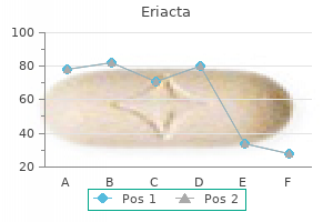
Management techniques vary: some therapies are chosen according to type of pain and underlying mechanism of action erectile dysfunction treatment miami buy 100 mg eriacta fast delivery, whereas others are selected on the basis of the diagnosis impotence at 19 purchase eriacta no prescription. Either way erectile dysfunction treatment pakistan buy 100mg eriacta otc, it is important to consider the mechanism of action erectile dysfunction klonopin buy genuine eriacta online, side effects erectile dysfunction doctor in pune cheap 100 mg eriacta free shipping, dosing, drug-drug interactions, and the individual patient in determining the appropriate therapies. When prescribing opioids, clinicians must also be vigilant for abuse and counsel patients on the appropriate use of the medication. In general, the use of a scheduled pain regimen is preferred over as-needed dosing. If pain is not controlled, opioids can be added to the regimen, with caution because of the potential side effects of sedation, respiratory depression, nausea, and vomiting. The use of opioids in mechanically ventilated patients can have good results, but with prolonged treatment, patients may develop dependence and will need to be weaned off them. Addressing Prescription Drug Abuse in the United States: Current Activities and Future Opportunities. Vital signs: overdoses of prescription opioid analgesics-United States, 1999-2008. A mechanism-based approach to pain pharmacotherapy: targeting pain modalities for optimal treatment efficacy. Epidemiology of chronic pain with psychological comorbidity: prevalence, risk, course, and prognosis. Efficacy of desipramine in painful diabetic neuropathy: a placebo-controlled trial. Amitriptyline relieves diabetic neuropathy pain in patients with normal or depressed mood. A review of its pharmacological properties and therapeutic use in chronic pain states. A comprehensive review of clinical trials on the efficacy and safety of drugs for the treatment of low back pain. Zonisamide in the treatment of painful diabetic neuropathy: a randomized, double-blind, placebo-controlled pilot study. Safety and effectiveness of topiramate for the management of painful diabetic peripheral neuropathy in an open-label extension study. Efficacy of interdisciplinary treatment for chronic nonmalignant pain patients in Japan. Incidence of constipation associated with long-acting opioid therapy: a comparative study. Transdermal fentanyl versus sustained release oral morphine in strong-opioid naive patients with chronic low back pain. Vital signs: variation among states in prescribing of opioid pain relievers and benzodiazepines- United States, 2012. Substance Abuse and Mental Health Services Administration, Center for Behavioral Health Statistics and Quality. Behavioral Health Coordinating Committee, Prescription Drug Abuse Subcommittee, U. Complications of long-term opioid therapy for management of chronic pain: the paradox of opioid-induced hyperalgesia. Mechanisms of hyperalgesia and morphine tolerance: a current view of their possible interactions. The role of ketamine in preventing fentanyl-induced hyperalgesia and subsequent acute morphine tolerance. The cyclooxygenase isozyme inhibitors parecoxib and paracetamol reduce central hyperalgesia in humans. Pain: moving from symptom control toward mechanismspecific pharmacologic management. The common successful scenario follows a typical path: an innovative novel procedure, supported by a single or very few successful case series, is proposed; it is followed by further case reports by interested surgeons; interest becomes more widespread; the procedure is gradually adopted into general surgical practice; and finally it becomes the "gold standard. Even procedures that become established may face rejection if in popular use they are shown to be associated with excessive risk or poor tolerability or are simply perceived as less effective than an alternative intervention. In the process, many patients will have been subjected to surgical interventions that have limited value. Although multiple mechanisms are necessary to dictate the fate of a particular intervention, an important role is played by key opinion leaders whose views remain unchallenged because of the lack of objective evidence to the contrary. This scenario was played out in the 20th century in the surgical management of trigeminal neuralgia. The notable limitations of pharmacologic treatment paved the way to the evolution of surgical methods ranging from division of the trigeminal nerve to alcohol injections, neurectomy, radiofrequency rhizotomy, glycerol rhizotomy, and balloon microcompression. First, vascular compression or contact cannot be the primary cause because (1) it is common in asymptomatic people9; (2) in up to 28% of patients with classic trigeminal neuralgia, no compression can be found10; and (3) trigeminal neuralgia does recur over the years in 25% of affected patients without evidence of a new compression. When the surgeon discusses the advantages and disadvantages of various procedures with the patient, results from comparative trials showing superiority of a given procedure over the others would be helpful; however, there are none. Observational studies, however, have been conducted with clear patient selection, careful data collection, independent assessors, and actuarial outcome presentation. However, it does not answer the question of whether in a particular case the chosen method is optimal. The currently available procedures are, without doubt, extraordinarily effective in treating trigeminal neuralgia. In a patient with refractory trigeminal neuralgia, the remarkable outcome of postoperative freedom of pain for several years is evidence enough. Unfortunately, only low-quality comparative studies to that effect have been published. They are mostly single-center retrospective assessments of outcomes from two or more procedures with inconstant follow-up (for reviews, see Pollock1; Tatli and associates11; and Zakrzewska and colleagues31). The uncertainty over the best method becomes even greater if the clinician wishes to establish which approach is best for subgoups of patients with trigeminal neuralgia associated with multiple sclerosis, trigeminal neuralgia with atypical features, and trigeminal neuralgia that recurs after an earlier operation. The failure of any of the comparative studies published so far to identify a superior treatment results from their inherent biases. The major bias concerns the difference between compared patient groups because of lack of randomization. The groups tend to differ in previous treatment received, duration of trigeminal neuralgia, associated disability, clinical features (especially those of atypical trigeminal neuralgia), and previous responses to therapy. In addition, because of the high attrition rate in long-term studies and the general tendency not to use an independent party for outcome assessment, the risk of overall bias is too high for conclusions. We carried out a systematic review of publications from 1980 to 2013 and found 14 procedures that were advocated by various authors. Of interest was that no intervention was superior to the rest in efficacy or safety. The relative similarity of benefit across all reported methods reveals the same problem as in the surgical management of trigeminal neuralgia: for the surgeon, the choice of the best method for the patient remains arbitrary. First, chronic pain is subjective, and its intensity notoriously fluctuates over time; some conditions are even known to go into long spontaneous remissions. Second, for chronic pain, there are many treatments easily accessed by patients, seemingly similar in outcome, which enhances the risk of bias from patients. Third, the placebo reaction is greater in chronic pain than in any other condition, except depression, and accounts for up to 44% of the treatment response. Comparison of a novel treatment with sham treatment will show whether the novel treatment has some efficacy in a given condition and, in most cases, the magnitude of the effect. In contrast, studies that lack randomization and control yield results applicable only to that particular study population, and generalizability is limited. With few exceptions, such studies fail to address the very essence of the matter: whether a particular treatment is superior to an alternative treatment or sham treatment. In cluster headache, both individual experts and the Task Force of the European Headache Society emphasize the need for adequately powered controlled trials before treatment recommendations can be formulated. For treatment of attacks, another multicenter study focused on the efficacy of sphenopalatine ganglion stimulation versus control for 15 minutes before use of acute medication. Although the results were negative, they reflected the practicalities of the study rather than its design. Unless the effect sizes are substantial, this would not be in the interest of therapeutic evolution. Rather, a further step would be a comparative trial between balloon microcompression of the gasserian ganglion or radiofrequency lesioning of the sphenopalatine/ trigeminal ganglion for both efficacy and cost effectiveness. As discussed previously, in trigeminal neuralgia little benefit is expected from any sham-controlled efficacy studies, now that case series in large patient populations have shown a substantial effect size. However, comparative studies involving subgroups of patients with trigeminal neuralgia are clearly needed and have significant potential in providing clinically meaningful information. This idea is based on reports of relative success both from posterior fossa exploration and balloon compression and radiofrequency rhizotomy. One should not be reluctant to conduct such studies because of the difficulties that they entail, but without them (or similar controlled trials), a large percentage of patients with trigeminal neuralgia remain in danger of receiving inferior care. Clearly, blind conditions may be impossible or must be compromised to an unsatisfactory degree. These issues, however, are not insurmountable, and alternative trial designs can be used to address them. They are appropriate when used to test early innovative ideas and when researchers gather data to allow sample size estimates for pivotal controlled trials. The mistake in the past was to assume that observational data per se suffice to prove a hypothesis, which is not the case. Scientifically robust methods to assess the value of a new intervention should be rigorously employed. Trials such as the ones just proposed would undoubtedly add to clinical practice but are costly and time consuming. They require multicenter and even international collaborations to be adequately powered, with added attention to standardization of surgical procedures and patient selection. Trigeminal neuralgia subtypes, in contrast, have not been rigorously assessed in this regard, and there are no reliable data to be used for treatment recommendations. The National Neurosurgery Quality and Outcomes Database and NeuroPoint Alliance: rationale, development, and implementation. Surgical management of trigeminal neuralgia: use and cost-effectiveness from an analysis of the Medicare Claims Database. A comparison of percutaneous radiofrequency trigeminal neurolysis and microvascular decompression of the trigeminal nerve for the treatment of tic doloureux. CyberKnife radiosurgical rhizotomy for the treatment of atypical trigeminal nerve pain. Practice patterns of in-hospital surgical treatment of trigeminal neuralgia from 1980 to 2010. The long-term outcome predictors of pure microvascular decompression for primary trigeminal neuralgia. Recovery of nerve conduction following microvascular decompression for trigeminal neuralgia. Operative findings and outcomes of microvascular decompression for trigeminal neuralgia in 35 patients affected by multiple sclerosis. Predictors of outcome in surgically managed patients with typical and atypical trigeminal neuralgia: comparison of results following microvascular decompression. Clinical features and surgical treatment of trigeminal neuralgia caused solely by venous compression. Micro-vascular decompression for primary trigeminal neuralgia (typical or atypical). Long term effectiveness on pain: prospective study with survival analysis in a consecutive series of 362 patients. Repeat posterior fossa exploration for patients with persistent or recurrent idiopathic trigeminal neuralgia. Comparison of surgical treatments for trigeminal neuralgia: re-evaluation of radiofrequency rhizotomy. Percutaneous stereotactic radiofrequency lesioning for trigeminal neuralgia: determination of minimum clinically important difference in pain improvement for patient-reported outcomes. Microvascular decompression for classic trigeminal neuralgia: determination of minimum clinically important difference in pain improvement for patientreported outcomes. Microvascular decompression versus percutaneous procedures for typical trigeminal neuralgia: personal experience. Patient reports of satisfaction after microvascular decompression and partial sensory rhizotomy for trigeminal neuralgia. Results from the use of glycerol injection, microvascular decompression, and rhizotomia. Comparison of percutaneous trigeminal ganglion compression and microvascular decompression for the management of trigeminal neuralgia. Fifteen years of Gamma Knife surgery for trigeminal neuralgia in the Journal of Neurosurgery: history of a revolution in functional neurosurgery. Microvascular decompression for trigeminal neuralgia in patients with multiple sclerosis. Percutaneous retrogasserian glycerol rhizotomy for patients with idiopathic trigeminal neuralgia: a prospective analysis of factors related to pain relief. Arthroscopic partial meniscectomy versus sham surgery for a degenerative meniscal tear. Safety and efficacy in deep brain stimulation for refractory cluster headache: a randomized, placebo-controlled, double-blind trial followed by an open extension. Gamma Knife surgery for recurrent or residual trigeminal neuralgia after a failed initial procedure. Effectiveness of repeat glycerol rhizotomy in treating recurrent trigeminal neuralgia. The plaques of demyelination that occur lead to hyperexcitability of exposed and potentially injured afferents, which results in afterdischarges large enough to cause a nonnociceptive signal being perceived as pain. There may also be genetic and/or myelin biologic predispositions, given that rare reports of genetic and familial associations do exist. Hemminki and colleagues constructed a nationwide neurological database from the Swedish multigenerational register on 0- to 69-year-old siblings that was linked to the hospital discharge register for the 15-year period 1987 to 2001.
Order genuine eriacta on line. How To Prepare the Erecaid for Use.
References
- Marsh C, Shinde S. Neurosurgery and the parturient anaesthesia-Tutorial of the week 253.
- Rosero E, Joshi GP. Total intravenous anesthesia: Present and future. ASA Monitor. August 2015; 79(8):10-57.
- Kjaergaard J, Hastrup SJ, Sogaard P, et al. Advanced quantitative echocardiography in arrhythmogenic right ventricular cardiomyopathy. J Am Soc Echocardiogr. 2007;20:27-35.
- Tamber MS, Klimo P, Jr, Mazzola CA, et al: Pediatric hydrocephalus: systematic literature review and evidence-based guidelines. Part 8: management of cerebrospinal fluid shunt infection. J Neurosurg Pediatr 14(Suppl 1):60-71, 2014.
- Gandaglia G, Fossati N, Stabile A, et al: Radical prostatectomy in men with oligometastatic prostate cancer: results of a single-institution series with long-term follow-up, Eur Urol 72(2):289n292, 2017.
- Kubo H, Nakayama K, Yanai M, et al. Anticoagulant therapy for idiopathic pulmonary fibrosis. Chest 2005;128:1475-82.
- Carter P, Presta L, Gorman CM, et al. Humanization of an anti-p185HER2 antibody for human cancer therapy. Proc Natl Acad Sci U S A 1992;89(10): 4285-4289.