

Katarzyna Jadwiga Macura, M.D., Ph.D.
- Assistant Director ICTR Imaging Translational Program
- Professor of Radiology and Radiological Science

https://www.hopkinsmedicine.org/profiles/results/directory/profile/0015445/katarzyna-macura
Following this arrhythmia education inc lopressor 25 mg fast delivery, a lichenoid dermatitis with variable psoriasiform hyperplasia occurs Direct immunofluorescence yields negative findings arrhythmia definition medical order genuine lopressor. Dapsone and alteration of the diet are also effective; topical steroids are not effective prehypertension and alcohol 50mg lopressor with amex. Skin biopsies reveal a dense lymphohistiocytic infiltrate arrhythmia general anesthesia discount lopressor 12.5mg mastercard, eosinophils in the papillary dermis blood pressure chart with age lopressor 100 mg on-line, and increased Langerhans cells. History will assist in making the diagnosis of atropic dermatitis, whereas biopsy may reveal findings diagnostic of eruptions. Systemic steroids are the treatment of choice and may result in long-term remission. The vulva, scrotum, and anal areas are common sites, although the genital and anal areas are seldom involved at the same time. The eruption may be papular, resembling lichen planus; and in other cases the patches are excoriated, slightly scaly or moist, and rarely, nodular. Persistent rubbing of the shins or upper back may result in dermal deposits of amyloid and the subsequent development of lichen or macular amyloidosis, respectively. Chronic scratching of a localized area may be a response to an inciting dermatitis; however, scratching of the localized site continues long after the original insult and becomes a habit. It may be associated with anxiety disorders and in depressed patients, with erectile dysfunction. This change, known as lichenification, may originate on seemingly normal skin or may develop on skin that is the site of another disease, such as atopic or allergic contact dermatitis or ringworm. It is important to stress the need for the patient to avoid scratching the areas involved if the sensation of itch is ameliorated. Recurrences are frequent, even after the most thorough treatment, and in some cases the clearance of one lesion will see the onset of another elsewhere. A high-potency steroid cream or ointment should be used initially but not indefinitely because of the potential for steroidinduced atrophy. Use of a steroid-containing tape to provide both occlusion and antiinflammatory effects may have benefit. Treatment can be shifted to the use of medium- to lower-strength topical steroid creams as the lesions resolve. Topical doxepin, capsaicin, or pimecrolimus cream or tacrolimus ointment provides significant antipruritic effects and is a good adjunctive therapy. Too superficial an injection invites the twin risks of epidermal and dermal atrophy and depigmentation, which may last for many months. The suspension should not be injected into infected lesions because it may cause abscess. Sorenson E et al: Successful use of a modified Goeckerman regimen in the treatment of generalized prurigo nodularis. The individual lesions are pea sized or larger, firm, and erythematous or brownish. Prurigo nodularis is one of the disorders in which the pruritus is characteristically paroxysmal: intermittent, unbearably severe and relieved only by scratching to the point of damaging the skin, usually inducing bleeding and often scarring. The histologic findings are those of compact hyperkeratosis, irregular acanthosis, multinucleated keratinocytes, and a perivascular mononuclear cell infiltrate in the dermis. Delusions of parasitosis, psychogenic (neurotic) excoriations, factitial dermatitis, and trichotillomania compose the major categories of psychodermatology. The differential diagnosis for these four disorders is twofold, requiring the exclusion of organic causes and the definition of a potential underlying psychological disorder. The initial treatment of choice for prurigo nodularis is intralesional or topical administration of steroids. Usually, superpotent topical products are required, but at times, lowerstrength preparations used with occlusion may be beneficial, as when administered as the "soak and smear" regimen. The use of steroids in tape (Cordran) and prolonged occlusion with semipermeable dressings, such as used for treating nonhealing wounds, can be useful in limited areas. The combination product containing calcipotriene and betamethasone dipropionate ointment, calcitriol ointment, or tacrolimus ointment applied topically twice daily may be therapeutic and steroid sparing. Managing dry skin with emollients and avoidance of soap, with administration of antihistamines, antidepressants, or anxiolytics, is of moderate benefit in allaying symptoms. Good results have been obtained with thalidomide, lenalidomide, pregabalin, and cyclosporine. With thalidomide, onset may be rapid or slow, and sedation may occur; initial dose is 100 mg/day, titered to the lowest dose required. Lenalidomide, an analog of thalidomide, has less problems with neuropathy but may cause myelosuppression, venous thrombosis, and Stevens-Johnson syndrome. Multimodal therapy, combining topical and systemic therapies, can improve results. In cases associated with renal failure, transepidermal elimination of degenerated collagen may be found. Some of the signs described here may become repetitive compulsions that impair normal life functions and may be manifestations of an obsessive-compulsive disorder. Self-injury by prolonged, compulsive repetitious acts may produce various mutilations, depending on the act and site of injury. Bumping of the head produces lacerations and contusions, which may be so severe as to produce cranial defects and life-threatening complications. Bulimia, with its self-induced vomiting, results in Russell sign-crusted papules on the dorsum of the dominant hand from cuts by the teeth. Clenching of the hand produces swelling and ecchymosis of the fingertips and subungual hemorrhage. The belief is so fixed that the patient often pick small pieces of epithelial debris from the skin and bring them to be examined, insisting that the offending parasite is contained in such material Samples of alleged parasites enclosed n assorted containers, paper tissue, or sandwiched between adhesive tape are so characteristic that it is referred to as the "matchbox" or "ziplock" sign. Intranasal formication, or a crawling sensation of the nasal mucosa, is common in this condition. Cutaneous findings may range from none to excoriations, prurigo nodularis, and frank ulcerations. Women are affected 2: 1 over men, often during middle or old age, although there is a bimodal peak, with some patients presenting in their 20s or 30s. Body dysmorphic disorder is a spectrum of disease; some severely affected patients are delusional, whereas others have more insight and are less functionally impaired. Monosymptomatic hypochondriacal disorder is a form of psychosis characterized by delusions regarding a particular hypochondriacal concern. In contrast to schizophrenia, there are no other mental deficits, such as auditory hallucination, loss of interpersonal skills, or presence of other inappropriate actions. Patients with monosymptomatic hypochondriacal psychosis often function appropriately in social settings, except for a single fixated belief that there is a serious problem with their skin or with other parts of their body. Self-inflicted lacerations may be of suicidal intent Lip licking produces increased salivation and thickening of the lips. Eventually, the perioral area becomes red and produces a distinctive picture resembl ng the exaggerated mouth makeup of a clown. Pressure produced by binding the waistline tightly with a cord will eventually lead to atrophy of the subcutaneous tissue. Psychopharmacologic agents, especially the newer atypical antipsychotic agents, and behavioral therapy alone or in combination with these agents are the treatments of choice. They include cocaine, alcohol, and amphetamine abuse; dementia and other neurologic conditions. A variety of medications, including gabapentin, antiparkinsonian and antihistaminic drugs, and corticosteroids, may also produce this condition. Some of these agents may produce cutaneous symptoms, particularly pruritus, which may contribute to the delusion. Initial steps should be directed at excluding a true infestation, such as scabies, or an organic cause. A thorough history, particularly in reference to therapeutic and recreational drug use. Many consider Morgellons disease simply to be another name for delusions of parasitosis. Patients complain of crawling, biting, burning, or other sensations that cause them to be intensely anxious. A skin biopsy is frequently performed, more to reassure the patient than to uncover occult skin disease. Once organic causes have been eliminated, the patient should be evaluated to determine the cause of the delusions. Schizophrenia, monosymptomatic hypochondriacal psychosis, psychotic depression, dementia, and depression with somatization are considerations in the differential diagnosis. Although referral to a psychiatrist may seem best, most frequently the patient will reject suggestions to seek psychiatric help. The dermatologist is cautioned against confronting the patient with the psychogenic nature of the disease. If pharmacologic treatment is undertaken, the patient may accept it if the medication is presented as one that will alter the perception of this bothersome sensation. Although this strategy has limited side effects, newer, atypical antipsychotic agents, such as risperidone (0. Many persons have unconscious compulsive habits of picking at themselves, and at times the tendency is so persistent and pronounced that excoriations of the skin result. The lesions are caused by picking, digging, or scraping and usually occur on parts readily accessible to the hands. These patients admit their actions induce the lesions but cannot control their behavior. Righthanded persons tend to produce lesions on their left side and left-handed persons on their right side. There is evidence of past healed lesions, usually with linear scars, or rounded hyperpigmented or hypopigmented lesions, in the area of the active excoriations. The organic differential diagnosis is vast and includes any condition that may manifest with excoriations. The most common psychopathologies associated with neurotic excoriations are depression, obsessive-compulsive disorder, and anxiety the treatment of choice is doxepin because of its antidepressant and antipruritic effects; doses are slowly increased to 100 mg or higher, if tolerated. Many alternatives to doxepin may be indicated, especially in hose affected by an obsessive-compulsive component. N-acetylcysteine 1200 to 3000 mg/day is safe and tolerable option, which is now being studied in many psychodermatologic conditions. Treatment is difficult, often requiring a combined psychiatric and pharmacologic intervention. An attempt should be made to identify specific conflicts or stressors preceding onset. The therapist should concentrate on systematic training directed at the behavioral reaction pattern. Malingering applies to the latter two cases, where material gain is the objective. This contrasts with the usual dermatitis artefacta patient, who has an unconscious goal of gaining attention and assuming the sick patient role. Most patients are adults in midlife, with women affected three times more often than men. These skin lesions are provoked by mechanical means or by the application or injection of chemical irritants and caustics. These patients often have a "hollow" history, unable to detail how the lesions appeared or evolved. The lesions are generally distributed on parts easily reached by the hands, tend to be linear and arranged regularly and symmetrically, and are rarely seen on the dominant hand. When chemicals are used, red streaks or guttate marks are often seen beneath the principal patch, where drops of the chemical have accidentally run or fallen on the skin. According to the manner of production, the lesions may be erythematous, vesicular, bullous, ulcerative, or gangrenous. The more common agents of destruction used are the fingernails, pointed instruments, hot metal, chemicals. At times the only sign may be the indefinitely delayed healing of an operative wound, which is purposely kept open by the patient. Tight cords or clothing tied around an arm or leg may produce factitious lymphedema, which may be mistaken for postphlebitic syndrome or nerve injury, as well as other forms of chronic lymphedema. Subcutaneous emphysema, manifesting as cutaneous crepitations, may be factitial in origin. Recurrent migratory subcutaneous emphysema involving the extremities, neck, chest, or face can be induced through injections of air into tissue with a needle and syringe. Circular pockets and bilateral involvement without physical findings, indicating contiguous spread from a single source, suggest a factitial origin. Puncturing the buccal mucosa through to facial skin with a needle and puffing out the cheeks can produce alarming t ne ne. Neck and shoulder crepitation is also a complication in manic patients that results from hyperventilation and breath holding the organic differential diagnosis depends on the cutaneous signs manifested, such as gas gangrene for patients with factitious subcutaneous emphysema and the various forms of lymphedema for factitious lymphedema. They tend to cause lesions that closely simulate known conditions, and they create an intricate, often fantastic story surrounding the problem. Parents may induce lesions on their child to gain attention, so-called Munchausen by proxy, which is really child abuse. Considerations for psychopathology in dermatitis artefacta include borderline personality disorders and psychosis. Occlusive dressings may be necessary to protect the lesions from ready access by the patient.
Fatal cases of hyperinfection occur in immunocompromised patients; the parasite load increases dramatically and can produce a fulminant illness arteria carotida externa cheap lopressor online mastercard. Widespread petechiae and purpura are helpful diagnostic signs of disseminated infection hypertension images buy 50 mg lopressor amex, and chronic urticaria is a possible presenting sign pulse pressure 32 order 12.5mg lopressor fast delivery. In one reported case pulse pressure 48 buy lopressor cheap, widespread follicular arrhythmia in child buy cheap lopressor, erythematous, dome shaped papules and pustules appeared on the patient within 24 hours of working under a house. Scraping the lesions revealed live and dead larvae of the free-living soil nematode Pelodera strongyloides. It is acquired from body contact with damp sand or earth that has been contaminated by the excreta of dogs and cats. The diagnosis is typically made clinically, although biopsy may sometimes demonstrate the organism, and even dermoscopy has been used. Criteria for successful therapy are relief of symptoms and cessation of tract extension, which usually occurs within 1 week. Both ivermectin and metronidazole have been used topically, as has thiabendazole, compounded as a 10% suspension or a 15% cream. Histopathologic examination of the skin swelling will demonstrate eosinophilic panniculitis. Eating raw flesh from the second intermediate host, most often freshwater fish, especially eel, in such preparations as sashimi and ceviche, allows humans to become the definitive host. As the larval cyst in the flesh Guinea worm disease is now limited to remote villages in several sub-Saharan African countries It is caused by Dracunculus medinensis and is contracted through drinking water that has been contaminated with infected water fleas in which Dracunculus is parasitic. In the stomach, the larvae penetrate into the mesentery, where they mature sexually in 10 weeks. The female worm then burrows to the cutaneous surface to deposit her larvae and thus causes the specific skin manifestations. As the worm approaches the surface, it may be felt as a cordlike thickening and forms an indurated cutaneous papule. When the parasite comes in contact with water, the worm rapidly discharges its larvae, which are ingested by water fleas (Cyclops), contaminating the water. It is characterized by lymphedema, with resulting hypertrophy of the skin and subcutaneous tissues, and by enlargement and deformity of the affected parts, usually the legs, scrotum, or labia majora. The onset of elephantiasis is characterized by recurrent attacks of acute lymphangitis in the affected part, associated with chills and fever (elephantoid fever) that last for several days to several weeks. After each attack, the swelling subsides only partially, and as recrudescences supervene, thickening and hypertrophy become increasingly pronounced. The overlying epidermis becomes stretched, thin, and shiny, and over years, leathery, insensitive, and verrucous or papillomatous from secondary pyogenic infection. In addition to involvement of the legs and scrotum, the scalp, vulva, penis, female breasts, and arms can be affected, either alone or in association with the other regions. When the legs are attacked, both are usually affected somewhat symmetrically, with the principal changes occurring on the posterior aspects above the ankles and on the dorsa of the feet. At first, the thickening may be slight and associated with edema that pits on pressure. When the scrotum is affected, it gradually reaches an enormous size, and the penis becomes hidden in it. Dracunculiasis may be prevented by boiling drinking water, providing safe drinking water through boreholes, or filtering the water through mesh fibers. Native treatment consists of gradually extracting the worm a little each day, taking care not to rupture it; otherwise, the larvae escape into the tissues and produce fulminating inflammation. Metronidazole, 500 mg/day, resolves the local inflammation and permits easier removal of the worm. Global eradication is within reach, and Guinea worm disease may become a historical footnote. A testicle may enlarge rapidly to the size of an apple and may be extremely painful. As a result of obstruction and dilation of the thoracic duct or some of its lower abdominal tributaries into the urinary tract, chyle appears in the urine, which assumes a milky appearance. Lobulated swellings of the inguinal and axillary glands, called varicose glands, are caused by obstructive varix and dilation of the lymphatic vessels. Filaria are transmitted person to person by the bites of a variety of mosquitoes of the Culex, Aedes, and Anopheles species. Microfilarial embryos may be seen as coiled, each in its own membrane near the posterior tip. It is important to realize that infestation by the filaria is often asymptomatic, and elephantiasis usually occurs only if hundreds of thousands of mosquito bites occur over a period of years, with episodes of intercurrent streptococcal lymphangitis. Filariasis was endemic in the considerable Samoan population of Hawaii for half a century, and only one case of elephantiasis has occurred among this group. The microfilariae should be sought on fresh coverslip films of blood (collected at night), urine, or other body fluid and examined with a low-power objective lens Calcified adult worms may be demonstrated on x-ray examination, and ultrasound can detect adult worms. At times, adult filariae are found in abscesses or in material taken for pathologic examination. Specific serologic tests and a simple card test for filarial antigen are available. The prognosis in regard to survival is good, but living becomes burdensome unless the condition is alleviated. Diethylcarbamazine, in increasing doses over a 14-day period, is the treatment of choice. This leads to long-term sterility of adult female worms and both are being studied to determine their place in the treatment of both bancroftian filariasis and onchocerciasis. In infected persons, the parasite develops slowly, and even 3 years can elapse between infection and appearance of symptoms, although the usual interval is 1 year. They typically last a few days and then subside, although recurrent swellings at the same site may eventually lead to a permanent, cystlike protuberance. These swellings may result from hypersensitivity to the adult worm or to materials elaborated by it. The filariae may be noticed subcutaneously in the fingers, breasts, eyelids, or submucosally under the conjunctivae. The worm may be in the anterior chamber of the eye, the myocardium, or other sites. It has a predilection for loose tissues such as the eye region, the frenum of the tongue, and the genitalia. The wanderings of the adult parasite may be noticed because of a tingling and creeping sensation. The death of the filaria in the skin may lead to the formation of fluctuant cystic lesions. Loiasis is widely distributed in West and Central Africa, where it is transmitted by the mango fly, Chrysops dimidia or Chrysops silacea. The observation of the worm under the conjunctiva, Calabar swellings, eosinophilia, and microfilariae in peripheral blood establish the diagnosis. This must be done quickly by seizing the worm with forceps and placing a suture under it ok fre been devised to remove the edematous subcutaneous tissue from the scrotum and breast. If a trip of over 1 month to areas with endemic Wuchereria bancrofti is planned and extensive exposure to mosquitoes is likely, taking diethylcarbamazine, 500 mg/day for 2 days each month, is recommended. Worms that are not securely and rapidly grasped may escape into the deeper tissues. Diethylcarbamazine kills both adults and microfilariae and is given in increasing doses for 21 days. In regions where onchocerciasis and loiasis both are endemic, and where ivermectin is used in a community-based elimination strategy for onchocerciasis, simultaneously infected patients with a high L. If ivermectin treatment of these patients is undertaken, proper monitoring and appropriate supportive treatment should be available in anticipation of this risk. Diethylcarbamazine is an effective chemopreventive therapy, using 300 mg/week in temporary residents of regions of Africa where L. The dermatitis is variable in appearance, probably related to chronicity of infection, age of the patient, geographic area where acquired, and relative immune responsiveness. In Central America, another manifestation of the acute phase of onchocerciasis is acute swelling of the face with erythema and itching, known as erisipela de la costa In Zaire and Central America, an acute urticarial eruption is seen the inflammation, which is accompanied by hyperpigmentation, is known as mal morado. As time passes, the dermatitis becomes chronic and remains papular; however, thickening, lichenification, and depigmentation occur. When the depigmentation is spotted, it is known as leopard skin; when the skin is thickened, it is called elephant skin. When local edema and thickened, wrinkled, dry dermatitic changes predominate, it is sometimes called lizard skin. In Saudi Arabia, Yemen, and East Africa, a localized type of onchocerciasis exists called sowda, Arabic for "black " It is characterized by localized, pruritic, asymmetric, usually darkly pigmented, chronic lichenified dermatitis of one leg or one body region. It is also known as the chronic hyperreactive type, and an association with antidefensin antibodies suggests a reason for this enhanced reactivity against the parasite. After a time, firm subcutaneous nodules, pea-sized or larger, develop on various sites of the body. In parts of Africa, where natives are wholly or nearly unclothed, the lesions occur on the trunk, axillae, groin, and perineum. In Central and South America, the head, especially the scalp, is the usual site of involvement. Firm, nontender lymphadenopathy is a common finding in patients with chronically infected onchocerciasis. In about 5% of affected persons, serious eye lesions arise late in the disease, gradually leading to blindness. Onchocerciasis is caused by Onchocerca volvulus, which is transmitted to humans by the bite of the black fly of the genus Simulium. Millions of the progeny then migrate back into the dermis and the aqueous humor of the eye. Onchocerciasis occurs in Africa on the west coast, in the Sahara, Sudan, and the Victoria Nile division, where it is known as river. In Central and South America, this disease can be found in Guatemala, Brazil, Venezuela, and southern Mexico. The presence of eosinophilia, skin lesions, and onchocercomas with ocular lesions is highly suggestive in endemic areas. Frequently, the microfilariae may be found in skin shavings or dermal lymph, even when no nodules are detectable. The scapular area is the favorite site for procuring specimens for examination by means of a skin snip. This is performed in the field or office by lifting the skin with an inserted needle and then clipping off a small, superficial portion of the skin with a sharp knife or scissors. The specimen is laid in a drop of normal saline solution on a slide with a coverslip and examined under the microscope. When patients with suspected onchocerciasis were given a single oral dose of 50 mg of diethylcarbamazine, a reaction consisting of edema, itching, fever, arthralgias, and exacerbation of pruritus was described as a positive Mazzotti test reaction, which supported the diagnosis of onchocerciasis. Community-based treatment protocols have the objective of eliminating onchocerciasis from endemic areas. Severe reactions, including neurologic disease, may occur in patients simultaneously infected with Loa loa. Doxycycline kills the intracellular symbiotic bacteria, Wolbachia, that appear to cause Mazzotti reactions and is being tested for long-term effects and determination of its place in the treatment of onchocerciasis and bancroftian filariasis. If there is eye involvement, prednisone, 1 mg/kg, should be started several days before treatment with ivermectin. Ng Nguyen D, et al: A systematic review of taeniasis, cysticercosis and trichinellosis in Vietnam. Rostami A, et al: Meat sources of infection for outbreaks of human trichinellosis. Veraldi S, et al: Treatment of hookworm-related cutaneous larva migrans with topical ivermectin. Ten percent of patients develop a bilateral, asymptomatic hand swelling that is especially prominent over the digits, as well as erythema along the perimeters of the palms and volar surfaces of the digits, which progresses to desquamation. In 20% of cases, a nonspecific macular or petechial eruption occurs, and splinter hemorrhages are occasionally present. In the average patient, eosinophilia begins about 1 week after infection and attains its height by the fourth week. The immunofluorescence antibody test has the greatest value in establishing early diagnosis. A 2-mm-thick slice of the muscle biopsy may be compressed between two glass slides to demonstrate the cysts. Insect repellents are effective in preventing disease transmission and are especially important during travel to areas where vector-borne disease is endemic. The American Academy of Pediatrics recommends concentrations of 30% or less in products intended for use in children.
Bacterial infection may be caused by streptococci high blood pressure medication and sperm quality order generic lopressor from india, s aphylococci hypertension nclex questions order generic lopressor on-line, Pseudomonas heart attack and vine cover order 50mg lopressor free shipping, or Corynebacterium arteria yugular externa buy lopressor 25 mg cheap. Streptococcal intertrigo favors the neck hypertension pregnancy order lopressor online, axillary, and inguinal folds of young children. There is a well-demarcated, fiery-red, moist, shiny surface and a foul smell, with an absence of satellite lesions. The differential diagnosis includes seborrheic dermatitis, intertriginous psoriasis, erythrasma, and if the groin lesions are fissured, Langerhans cell histiocytosis. Separating the apposing skin surfaces with gauze or InterDry Ag textile is helpful. The latter has an antimicrobial silver complex impregnated within the fabric that when placed in the folded area not only wicks away moisture, but also retains the activity against fungi and bacteria for up to 5 days. Botulinum toxin type A has been used to dry out areas predisposed to recurrent disease. Low-potency topical corticosteroids and topical tacrolimus are helpful to reduce inflammation, but these should always be used in conjunction with a topical antifungal or antimicrobial agent. Histologic examination generally demonstrates keratin pits lined by small cocci as well as filamentous bacteria. Topical erythromycin, mupirocin, or clindamycin is curative in pitted keratolysis. Miconazole or clotrimazole cream and Whitfield ointment are effective alternatives. An Bras Dermatol 2016; 91: 106 Pranteda G, et al: Pitted keratolysis, erythromycin, and hyperhidrosis Dermatol Ther 2014; 27: 101. Vascular gangrene, purpura fulminans, and diabetic gangrene are covered in Chapter 35 and necrotizing fasciitis earlier in this chapter. Onset is usually sudden and is characterized by a chill, a rise in temperature, marked prostration, and severe local pain. Gas bubbles (chiefly hydrogen) produced by the infection cause crepitation when the area is palpated. Gas gangrene is caused by a variety of Clostridium species, most frequently Clostridium perfringens, C. Clostridium spores are resistant to skin sterilization chemicals; if injecting a site that is being soiled by stool incontinence, a mechanical wash before the sterile procedure, followed by an occlusive sterile dressing, is recommended. This nonclostridial myositis may be clinically similar, but with delayed onset (several days). The purulent exudate has a foul odor, and gram-positive cocci in chains are present. It is important to distinguish these two entities, because involved muscle may recover in nonclostridial myositis, and debridement may safely be limited to removal of grossly necrotic muscle. Infections with both clostridial and nonclostridial organisms such as Streptococcus faecalis, S. Chronic Undermining Burrowing Ulcers (Meleney Gangrene) Chronic burrowing ulcer was first described by Meleney as postoperative progressive bacterial synergetic gangrene. It usually follows drainage of peritoneal abscess, lung abscess, or chronic empyema. After 1 or 2 weeks, the wound markings or retention suture holes assume a carbunculoid appearance, finally differentiating into three skin zones: outer, bright red; middle, dusky purple; and inner, gangrenous with a central area of granulation tissue. In Meleney postoperative progressive gangrene, the essential organism is a microaerophilic, nonhemolytic streptococcus (peptostreptococcus) in the spreading periphery of the lesion, associated with S. This disease is differentiated from ecthyma gangrenosum, which begins as vesicles, rapidly progressing to pustulation and gangrenous ulceration in debilitated patients, and is caused by P. Pyoderma gangrenosum occurs in a different setting, lacks the bacterial findings, and does not respond to antibiotic therapy. In polymicrobial infections imipenem or meropenem should be given as adjunctive therapy. Infections are seen most often in the cervicofacial area but also on the abdominal region, thoracic area, or pelvis. Diabetic and immunosuppressed patients and alcoholics with poor dental hygiene are particularly at risk. The lesions begin as firm nodules or plaques and develop draining sinuses Grains or sulfur granu es may be present in the exudate, as n fungal mycetomas. In the cervicofacial region, the infection is known as lumpy jaw the underlying bone may be involved with periostitis or osteomyelitis. Extension of the infection into the abdominal wall may produce draining sinuses on the abdominal skin. The condition is often clinically misdiagnosed as a malignancy; the histologic appearance of the characteristic granules allows diagnosis. Eosinophilic clubs composed of immunoglobulin are seen at the periphery of the granule (Splendore-Hoeppli phenomenon). Other effective medications have been ampicillin, erythromycin, tetracyclines, ceftriaxone, and clindamycin. Takazawa T, et al: A case of acute onset postoperative gas gangrene caused by Clostridium perfringens. This is usually considered a form of necrotizing fasciitis because it spreads along fascial planes. Peak incidence is between ages 20 and 50, although cases have been reported in children. Diabetes mellitus, obesity, poor personal hygiene, long-standing oral corticosteroid therapy, and chronic alcoholism are predisposing factors. Culture for aerobic and anaerobic organisms should be carried out, and appropriate antibiotics started; surgical debridement and general support should be instituted. Primary cutaneous disease also occurs in healthy individuals in the form of a draining abscess or lymphangitic nodules after a cutaneous injury Nocardia asteroides is usually responsible for the disseminated form of nocardiosis. A prick by a thorn or briar, other penetrating injury, or an insect bite or sting may be the inciting event. Some are branched, but filaments tend to be shorter and more fragmentary than those of Actinomyces. On Sabouraud dextrose agar, without antibacterial additives, there are creamy or moist, white colonies, which later become chalky and orange colored. They are usually on the buttocks and extremities and are often grouped closely together. Ecthyma gangrenosum occurs in debilitated persons who may be suffering from leukemia, in the severely burned patient, in pancytopenia or neutropenia, or in patients with a functional neutrophilic defect, terminal carcinoma, or other severe chronic disease. Healthy infants may develop lesions in the perineal area after antibiotic therapy in conjunction with maceration of the diaper area the classic vesicle suggests the diagnosis. The contents of the vesicles or hemorrhagic pustules will show gram-negative bacilli on Gram staining, and cultures will be positive for P. Because this is usually a manifestation of sepsis, the blood culture will also show P. Although ecthyma gangrenosum is classically associated with P aeruginosa infection, similar hemorrhagic pustules may occur from a variety of other gram-negative organisms. Sheffer S, et al: Lymphocutaneous nocardiosis caused by Nocardia brasiliensis in an immunocompetent elderly woman. Minocycline is an alternative Linezolid is active, but potential adverse effects limit its use. As the water temperature rises, free chlorine levels fall, even though total chlorine levels appear adequate. Soaking the affected finger in a 1% acetic acid solution twice a day has been found to be helpful. Trimming the onycholytic nail plate, followed by application of Neospor n solution twice a day, is also effective. The addition of granulocytemacrophage colony-stimulating factor to stimulate both proliferation and differentiation of myeloid precursors is an adjunct in a patient with myelodysplasia or treatment-induced neutropenia. Patients have a poorer prognosis if there are multiple lesions, if there is a delay in diagnosis and institution of appropriate therapy, and if neutropenia does not resolve by the end of a course of antibiotics. Instrumentation or catheterization increases the risk of this infection Other lesions also seen with Pseudomonas septicemia include sharply demarcated areas of cellulitis, macules, papules, plaques, and nodules, characteristically found on the trunk. Pseudomonas mesophilica, Burkholderia cepacia, Citrobacter freundii, and Stenotrophomonas maltophilia may also produce such skin lesions in immunocompromised individuals. With increasing inflammation and maceration, dermatophytosis may progress to dermatophytosis complex, in which many types of gram negative organisms may be recovered as it becomes more difficult to culture dermatophytes. Prolonged immersion may also cause hydration and maceration of the interdigital spaces, with overgrowth of gram-negative organisms. Culture of these subcutaneous abscesses will reveal Pseudomonas or other gram-negative bacteria, which likely originate in the macerated toe webs. However, once the scaling and peeling progress to white maceration, soggy scaling, bad odor, edema, and fissuring, treatment must also include topical antibiotics or acetic acid compresses. Full-blown gram-negative toe web infection with widespread denudation and erythema, purulence, and edema requires systemic antibiotics. One case occurred limited to the hand and wrist occluded under colonized rubber gloves. Most lesions occur on the sides of the trunk, axillae, breasts, buttocks, and proximal extremities. Associated complaints may include earache, sore throat, headache, fever, and malaise. Rarely, systemic infection may result; breast abscess and bacteremia have been reported. Large community outbreaks have occurred associated with public pools, and 27 employees of a cardboard manufacturing facility who were exposed to wet work developed Pseudomonas folliculitis of the extremities as an occupational disorder. Aeromonas hydrophilia was found to be responsible for a clinically similar folliculitis that affected two siblings playing in an inflatable swimming pool. In patients with fever, constitutional symptoms, or prolonged disease, a third-generation oral cephalosporin or a fluoroquinolone such as ciprofloxacin or ofloxacin may be useful. Preventive measures have been water filtration, automatic chlorination to maintain a free chlorine level of 1 ppm, maintenance of water at pH 7. Pseudomonas hot foot syndrome was reported in a group of 40 children who developed painful, erythematous plantar nodules or pustules after wading in a community pool whose floor was coated with abrasive grit. One biopsy showed neutrophilic eccrine hidradenitis; another revealed dermal microabscesses. If the patient is a swimmer or has diabetes, acetic acid compresses for 1 or 2 days before surgery may prevent this complication. External otitis must be distinguished from allergic contact dermatitis due to neomycin in Cortisporin otic suspension. Allergic contact dermatitis produces severe pruritus, although tenderness may also be noted. Dermatitis may extend down the side of the cheek in a pattern suggesting drainage of the suspension. The swelling, pain, and erythema are more pronounced, with purulence and a foul odor. Lastly, commercial ear piercing of the upper ear cartilage may lead to infection with Pseudomonas, with resulting cosmetic deformity t ne ne. Local application of antipseudomonal and antiinflammatory Cortisporin otic solution or suspension, or 2% acetic acid compresses with topical corticosteroids, will help clear this infection. In patients with otorrhea or pus emanating from the canal, if the symptoms have been present for 1 week or more, or if diabetes or an immunologic defect is present, cleansing the canal, visualizing the tympanic membrane for perforation, and other precautions will be most readily handled by an otolaryngology consultation. Application of otic Domeboro solution after swimming will help prevent recurrence. They differ from gram-negative infection in patients with acne in that the site of Pseudomonas colonization is the external ear, and topical therapy alone to the face and ears is sufficient for cure. Also, an outbreak of gram-negative pustular dermatitis on the legs, arms, torso, and buttocks occurred in a group of college students who hosted a mud-wrestling social event. Biscaye S, et al: Ecthyma gangrenosum, a skin manifestation of Pseudomonas aeruginosa sepsis in a previously healthy child. There is defective intracellular digestion of the bacteria once they have been phagocytosed. The granulomas may arise as masslike lesions or nodules, abscesses, or ulcerations. They favor the perineum but also affect the abdominal wall, thorax, extremities, and axilla. Histologically, foamy eosinophilic Hansemann macrophages contain calcified, concentrically laminated, intracytoplasmic bodies (Michaelis-Gutmann). Successful treatment of malacoplakia depends on the isolated organism; a fluoroquinolone such as ciprofloxacin or ofloxacin typically is useful. Coates M, et al: A case of cutaneous malakoplakia in the head and neck region and review of the literature. Am J Otolaryngol 2009; 30: 101 Flann S et al: Cutaneous malakoplakia in an abdominal skin fold J Am Acad Dermatol 2010; 62: 896.
Atypical parakeratosis is often associated with the presence of a concomitant pulse pressure norms purchase 100 mg lopressor with amex, more serious lesion and such specimens warrant careful inspection to ensure a high-grade lesion is not overlooked hypertension untreated generic lopressor 100mg on line. A small cluster of cells with high nuclear to cytoplasmic (N/C) ratios and irregularly distributed nuclei arrhythmia beta blocker buy 100mg lopressor mastercard. The cells have dense blood pressure medication diuretic buy lopressor 50mg low price, orange-s taining cytoplasm and could represent either reactive or dysplastic changes blood pressure chart 40 year old male lopressor 12.5mg discount. The cells are pleomorphic and have high nuclear to cytoplasmic (N/C) ratios and nuclear membrane irregularities. A squamous cell carcinoma cannot be excluded but would be more likely to have a background of necrosis or "clinging diathesis" (Pap stain). The cells have high nuclear to cytoplasmic (N/C) ratios, enlarged nuclei, and irregular nuclear contours (Pap stain). The cells have high nuclear to cytoplasmic (N/C) ratios and marked variation in nuclear size (Pap stain). I ndividually dispersed cells are inevitably present and intact fragments of carcinoma may or may not be seen. The cells have variable sizes and shapes, often with irregular cytoplasmic projections. The nuclei are often pyknotic (small and ink black), resulting in paradoxically low N /C ratios. I rregularly shaped pale areas or "ghost nuclei" may be seen, which represent poorly stained nuclei. Highly atypical single cells in a background of diathesis (tumor necrosis and associated inflammation). The surgical follow-up was invasive keratinizing squamous cell carcinoma (Pap stain). Note how deeply pink the cytoplasm of the keratinized cells stains, an indication of atypical keratin production (Pap stain). Keratinizing carcinoma cells often have rigid, irregular cytoplasmic projections not seen in benign processes. In this case, the malignant cell has the appearance of a tadpole ("tadpole cell"). Atypical nuclei may or may not be identifiable, but their presence can contribute to a definitive diagnosis (Pap stain). Multinucleated Cell Pattern Multinucleated cells may appear in Pap test specimens and can be striking in their appearance. While the varied morphologies help distinguish between most entities, some entities are similar in appearance and must be separated through the identification of subtle features. The nuclei are slightly enlarged but maintain their round shape and regular contours. The nuclei are usually uniform within a given cell but may become crowded and aggregate together. The specimen usually contains abundant reactive endocervical cells in the background with a range of appearances, including many that are more easily recognized as reactive. In this case, the foamy cytoplasm and lack of nuclear molding and "ground glass" chromatin both support an endocervical cell origin (Pap stain). Radiation may induce longstanding atypia in squamous cells in subsequent Pap test specimens, and these changes may persist well beyond the duration of the therapy itself. While these cells have enlarged nuclei, they also have a corresponding increase in the amount of cytoplasm, resulting in a nearly normal N /C ratio. The cytoplasmic vacuoles are well demarcated and may displace the nucleus, causing a signet ring appearance. N uclear changes include nuclear vacuolization, multinucleation, hyperchromasia, and wrinkling of nuclear membranes. The cells are enlarged but have abundant cytoplasm, which contains small vacuoles. A fragment of squamous cells, some of which are multinucleated, have anisonucleosis and coarse chromatin. Despite the increased nuclear sizes, the cells are also enlarged, contributing to normal nuclear to cytoplasmic (N/C) ratios. The cells in this fragment are enlarged and have enlarged nuclei, but the streaming nature is reminiscent of reparative change. The patient had a recent history of radiation therapy, which is likely responsible for these changes (Pap stain). This cell has an enlarged, hypochromatic nucleus with smooth nuclear contours and abundant cytoplasm (Pap stain). While their presence at one time was thought to indicate a threatened pregnancy the presence of, syncytiotrophoblasts is now understood to be incidental. The main differential diagnosis is multinucleated giant cells (histiocytes), which are more commonly seen in Pap test specimens and also have no known clinical significance. This large, multinucleated cell has a short, wide tail ("cytoplasmic extension") and dense cytoplasm. The nuclei tend to concentrate in the center of the cell, in contrast to the random and more peripheral distribution of nuclei seen in multinucleated giant cell histiocytes (Pap stain). The cells have small nucleoli, which can also be seen in syncytiotrophoblasts (Pap stain). These cells have numerous, uniform nucleoli which are randomly distributed throughout the cell, with some nuclei present at the periphery of the cell (Pap stain). While the infected cell (center) is multinucleated with nuclear molding, the chromatin does not have the typical "ground glass" appearance Instead, the nuclei contain Cowdry bodies (large intranuclear inclusions) (Pap stain). Key Features of Low-Grade Squamous Intraepithelial Lesion Nuclei are enlarged and at least 3 times the size of a normal intermediate cell nucleus. The presence of a sharp perinuclear cytoplasmic clearing (perinuclear halo) is characteristic. When seen, the most common component present is the epithelial component, which has the appearance of a high-grade carcinoma. When present, the mesenchymal cells can have diverse forms and often have enlarged nuclei with complex shapes, sometimes with multinucleation. Dispersed Atypical Cell Pattern the dispersed atypical cell pa ern describes the presence of an unusual appearing population of cells in the Pap test that exists as singly dispersed cells. A dispersed population of cells causes diagnostic difficulties because their true nature may only be determined at high magnification and also because the cells may be difficult to identify if they are present in low numbers in a specimen. S uperficial cells are deceased in number and may not be present at all, while intermediate and parabasal cells predominate. Parabasal cells can be seen in tissue fragments and/or as single, dispersed cells. The benign parabasal cells can have increased N /C ratios, hyperchromasia, and mild nuclear contour irregularities. The nucleus should be at least 3 times the size of a normal intermediate cell nucleus and contain other forms of nuclear atypia (hyperchromasia, nuclear contour irregularities, and/or bi-or multinucleation). This mature squamous cell has a markedly enlarged hyperchromatic nucleus (more than three times the size of the nuclei of the surrounding intermediate cells). Several cells have enlarged and hyperchromatic nuclei when compared with the background intermediate cells. I n addition to an increase in nuclear size, the nucleus should contain at least mild hyperchromasia or nuclear border irregularities. They have been given the name "litigation cells" because they are easily overlooked (due to their singly dispersed nature and because only rare cells may be present in a specimen). The four cells in this group have high nuclear to cytoplasmic ratios, hyperchromasia, and nuclear membrane irregularities. The cytoplasm is dense, with small "windows" between the cells, indicating that these may be metaplastic cells. The engulfed cell is markedly atypical with orangeophilic cytoplasm and an "ink black" hyperchromatic nucleus (Pap stain). This single cell in the center of field has an elevated nuclear to cytoplasmic (N/C) ratio and an irregularly shaped nucleus. Rare squamous cells with high nuclear to cytoplasmic ratios, hyperchromasia, and irregular nuclear contours are seen in a background of acute inflammation (Pap stain). The cell has a prominent nucleolus, dark chromatin, and a nuclear to cytoplasmic (N/C) ratio of around 0. Melanoma the cytologic features of melanoma seen on the Pap test are similar to those of melanoma in other sites. The neoplastic cells are enlarged and have round eccentric nuclei with regular borders. Melanin pigment may be seen in the cytoplasm and allow for a definitive diagnosis. The presence of binucleation, intranuclear inclusions, and/or prominent nucleoli may also suggest the possibility of melanoma. Malignant melanomas are more commonly metastatic rather than primary to the gynecologic tract; when metastatic melanoma is present, a history is usually known. Melanoma is determined to arise from the gynecologic tract in 5-10% of the cases encountered on Pap test specimens. I f cellularity is sufficient, an additional preparation can be made for confirmatory immunostains using melanoma-associated markers. The cells have abundant, granular cytoplasm, prominent nucleoli, and eccentrically placed nuclei. These features are suggestive of melanoma even in the absence of melanin pigment (Pap stain). A single, large binucleate cell with abundant cytoplasm is in the center of the field. The field also contains several much smaller melanoma cells with minimal cytoplasm; note that the chromatin pattern in these cells matches that of the two nuclei in the much larger cell (Pap stain). Small Cell Carcinoma S mall cell carcinoma of the cervix is a rare gynecologic malignancy and is morphologically similar to small cell carcinoma of the lung and other sites. I n liquid-based preparations, the visibility of background necrosis is reduced, though tumor necrosis it may be seen primarily as "clinging diathesis. Despite the name, the cells can look deceivingly large on cytologic preparations; the near-absence of cytoplasm is one clue to the diagnosis. Degenerated blood and granular debris (diathesis) is seen in the background and clinging to the neoplastic cells (Pap stain). Perinuclear Halo Pattern A cervical Pap test specimen may contain cells with a distinctive pale area surrounding the nucleus ("perinuclear halo" or "perinuclear clearing") that is sharply delineated from the rest of the cytoplasm by a crisp cytoplasmic border. S uperficial and intermediate squamous cells may occasionally have folded cytoplasm that gives the appearance of a perinuclear halo ("false halos"). When well developed, the perinuclear halo is characteristic and appears as an optically clear perinuclear area surrounded by a crisp cytoplasmic interface. While the cytoplasm appears pale in some areas, perinuclear "halos" are not well defined for these cells to be considered true koilocytes. More specific features include an irregularly shaped halo (rather than round) and a thickened interface between the cleared area and the rest of the cytoplasm (Pap stain). Other features of nuclear atypia are present: enlargement, irregular nuclear contours, hyperchromasia, and binucleation (Pap stain). Note the peripheral thickening of the cytoplasm, which is characteristic of koilocytes (Pap stain). A separate field containing a fragment of dysplastic cells in which nuclear enlargement was the primary diagnostic feature (Pap stain). Note that the cytoplasm is clear around the nuclei and thickened at the periphery. This intermediate cell has a very large nucleus that is also dark and irregularly shaped. This superficial cell has enlarged nuclei, binucleation, and a well-defined perinuclear halo. The perinuclear halo is poorly developed and has an indistinct border with the rest of the cytoplasm. Careful examination of the rest of the specimen may reveal better developed koilocytes (Pap stain). This small fragment of mature squamous cells demonstrates nuclear enlargement, perinuclear halos, and hyperchromasia (Pap stain). They are classically associated with Trichomonas infection, though Trichomonas organisms may not be seen along with inflammatory halos, either due to the nonspecific nature of these halos or lack of organisms due to clearance or recent treatment. Unlike the halos seen in navicular cells and koilocytes, inflammatory halos are more diffusely and uniformly seen through out the squamous cells in a specimen and do not have thickened borders. In addition to being small, the halos are also uniform and round and have indistinct borders with the rest of the cytoplasm. Although they are commonly known as "Trichomonas halos" (or "Trich halos"), they can sometimes be seen in other reactive conditions (Pap stain). The background contains numerous acute inflammatory cells; these findings together should prompt a careful search for Trichomonas organisms (Pap stain). Koilocytes tend to have optically clear halos and some form of nuclear atypia; navicular cell nuclei may have reactive changes but not the atypical nuclear features seen in koilocytes (enlarged, hyperchromatic, "raisinoid" nuclei). This suggests against these cells being koilocytes, with the accumulation of cytoplasmic glycogen being the most likely cause of these "halos" (Pap stain). The cell in the center of the field has a large, round perinuclear vacuole with a delicate interface with the rest of the cytoplasm. The faint yellow coloring inside this vacuole is not always identified but is associated with glycogen accumulation (Pap stain). Certain subtypes have been designated as "high-risk" based on whether a particular subtype has been associated with carcinogenesis. Eosinophilic (red on Pap stain) cytoplasmic granules, when seen, may aid in identification.
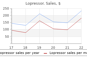
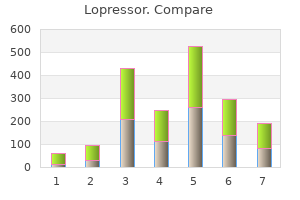
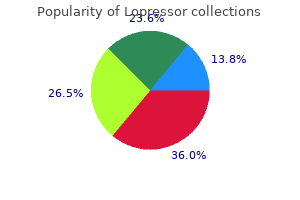
Purchase lopressor cheap online. how to lower high blood pressure 120/80 In 9 Minutes.
References
- Yohannan MD, Higgy KE, Al-Mashhadani SA, Santhosh- Kumar CR. Thrombocytosis. Etiologic analysis of 663 patients. Clin Pediatr. 1994;33:340-3.
- National Center for Biotechnology Information. dbSNP database. www.ncbi.nlm.nih.gov/snp Date last accessed: July 3, 2012.
- Amin MB, Tamboli P, Merchant SH, et al. Micropapillary component in lung adenocarcinoma: a distinctive histologic feature with possible prognostic significance. Am J Surg Pathol 2002;26(3):358-64.
- Norton P, Brubaker L: Urinary incontinence in women, Lancet 367(9504):57n67, 2006.
- Buch AN, Xue Z, Gevorkian NN, et al. Comparison of outcomes between bare metal stents and drug-eluting stents for percutaneous revascularization of internal mammary grafts. Am J Cardiol. 2006;98(6):722-724.
- Modlin IM, Lye KD, Kidd M. A 5-decade analysis of 13,715 carcinoid tumors. Cancer 2003;97:934.
- Gross M, Esterly JR, Earle RH. Pulmonary alterations in systemic lupus erythematosus. Am Rev Respir Dis 1972;105(4):572-7.