

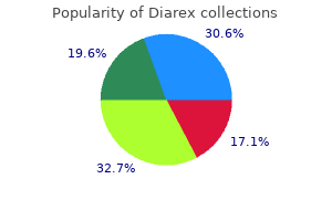
"Discount diarex 30 caps on line, lymphocytic gastritis definition".
N. Givess, M.A., M.D.
Associate Professor, New York University Long Island School of Medicine
Cautiously aspirate any blood or urine in the retropubic space gastritis b12 discount diarex 30caps line, but leave the area unexplored gastritis in english language diarex 30 caps with visa, as uncontrollable bleeding can result gastritis diet ����������� generic diarex 30 caps with amex. Open the peritoneum gastritis diet 360 discount diarex 30caps with mastercard, inspect the site of the rupture, and aspirate the fluid in the peritoneal cavity. Introduce a Foley catheter into the bladder through the urethra and then suture the tear with two layers of seromuscular stitches of 0 absorbable suture. It may be difficult to find but, if it is clearly visible, close it from within with 2/0 absorbable suture and insert a suprapubic catheter. The patient with a suprapubic catheter may start passing urine during this period; if so, remove the catheter. Referred abdominal pain Gastrointestinal obstruction, perforation and strangulation are important conditions which usually present with abdominal pain, although pain may also be referred. The location of referred abdominal pain is based on the embryological origin of the affected organ, while the location of peritoneal irritation depends on the anatomical position of the diseased organ. In cases where the diagnosis is not clear, repeated physical examination at frequent intervals will often clarify the need for surgery. Surgical exploration the treatment of many acute abdominal conditions includes surgical abdominal exploration. If surgery is indicated, do not avoid it in vulnerable patients including the young, old or pregnant. Use the midline incision which is simple, does not cause much bleeding, can be performed rapidly, closed quickly and extended easily. The midline laparotomy incision is described in Unit 6: Laparotomy and Abdominal Trauma. The gridiron incision for appendectomy is described on page 711 and the groin incision for hernia in Unit 8: Abdominal Wall Hernia. The surgical practitioner at the district hospital who can perform these three incisions can successfully manage most acute abdominal conditions. Findings that are important indications for surgery, are: Abdominal tenderness, suggesting inflammation of an underlying organ Rebound abdominal tenderness elicited by percussion, which confirms peritoneal irritation Involuntary contraction of the abdominal wall, a sign of peritoneal irritation, which presents as local guarding or generalized rigidity. Physical examination the history and physical examination are crucial to determine the most likely causes of an acute abdomen. The precise location of abdominal pain and 71 Surgical Care at the District Hospital 7 tenderness helps the practitioner to make a differential diagnosis. Although there are many acute abdominal conditions, only a few causes are common at any facility. Inflammatory bowel disease and colonic cancers are unusual at the district hospital while trauma, hernia and bowel obstruction are common. When doing a physical examination: Determine the vital signs Rapid respiration may indicate pneumonia Tachycardia and hypotension indicate patient decompensation Temperature is elevated in gastrointestinal perforation and normal in gastrointestinal obstruction Look for abdominal distension Percuss to differentiate gas from liquid Palpate the abdomen Start away from the site of tenderness Check for masses or tumours Determine the site of maximum tenderness Check for abdominal rigidity Listen for bowel sounds Absence is a sign of peritonitis or ileus High pitched tinkling indicates obstruction Always examine: Groin for incarcerated hernia Rectum for signs of trauma, abscess, obstruction Vagina for pelvic abscess, ectopic pregnancy, distended pouch of Douglas. Attention to hydration with intravenous fluid resuscitation is vital in all patients with intestinal obstruction. If the obstruction is not resolved, either by non-operative or operative treatment, intestinal gangrene or perforation will occur and lead to peritonitis. Bowel obstruction presents with: Abdominal pain, which may be colicky Vomiting Obstipation (absence of bowel movements and flatus) Abdominal distension. Bowel obstruction is a clinical diagnosis but it is greatly aided by plain erect and supine abdominal X-rays. The normal small bowel does not contain air and is therefore not visualized on X-ray. Distended loops of small bowel with air fluid levels are diagnostic of obstruction. Valvule coniventes cross the entire small bowel lumen and, when seen on X-ray, also indicate that the obstruction is intestinal. However, an obstruction caused by an underlying problem like abdominal abscess or generalized peritonitis will require surgery. The resuscitation procedure in non-operative management prepares the patient for surgery if it becomes necessary.
I am also grateful to Lauren Kerzin-Storrar and Tara Clancy for writing chapter 3 and to Dr Bronwyn Kerr for contributing to chapter 11 gastritis unusual symptoms proven 30caps diarex. Numerous colleagues have provided illustrations and are acknowledged throughout the book gastritis diet zinc purchase 30 caps diarex overnight delivery. In particular gastritis diet nhs purchase diarex 30 caps on-line, I would like to thank Professor Dian Donnai gastritis diet kolesterol diarex 30caps generic, Dr Lorraine Gaunt and Dr Sylvia Rimmer who have provided many illustrations for this as well as previous editions, and to Helena Elliott who has prepared most of the cytogenetic pictures incorporated into this new edition. I am also very grateful to the families who allowed me to publish the clinical photographs that are included in this book to aid syndrome recognition. Helen M Kingston vii this Page Intentionally Left Blank 1 Clinical genetic services Development of medical genetics the speciality of medical genetics is concerned with the study of human biological variation and its relationship to health and disease. It encompasses mechanisms of inheritance, cytogenetics, molecular genetics and biochemical genetics as well as formal, statistical and population genetics. Clinical genetics is the branch of the specialty involved with the diagnosis and management of genetic disorders affecting individuals and their families. Some of the disorders dealt with in these early clinics were ones that are seldom referred today, such as skin colour, eye colour, twinning and rhesus haemolytic disease. Other referrals were very similar to those being seen today namely, mental retardation, neural tube defects and Huntington disease. Prior to the inception of these clinics, the patterns of dominant and recessive inheritance, described by Mendel in 1865, were recognised in human disorders. Autosomal recessive inheritance of alkaptonuria had been recognised in 1902 by Archibald Garrod, who also introduced the term "inborn errors of metabolism". In 1908, the HardyWeinberg principle of population genetics was delineated and remains the basis of calculating carrier frequencies for autosomal recessive disorders. The term, "genetic counselling" was introduced by Sheldon Reed, whose definition of the process is given later in this chapter. The correct chromosome number in humans was not established until 1956 and the association between trisomy 21 and Down syndrome was reported in 1959. These advances have led to the mapping and isolation of many genes and subsequent mutation analysis. Enormous advances in molecular biology techniques have resulted in publication of the draft sequence of the human genome in 2000. As a result of these scientific discoveries and developments, clinical geneticists are able to use chromosomal analysis and molecular genetic tests to diagnose many genetic disorders. As more environmental diseases are successfully controlled those that are wholly or partly genetically determined are becoming more important. Despite a general fall in the perinatal mortality rate, the incidence of lethal malformations in newborn infants remains constant. Between 2 and 5% of all liveborn infants have genetic disorders or congenital malformations. These disorders have been estimated to account for one third of admissions to paediatric wards, and they contribute appreciably to perinatal and childhood mortality. Though diseases of wholly genetic origin are individually rare, they are numerous (several thousand single gene disorders are described) and are therefore important. Some are amenable to treatment but many are not, so that the emphasis is often placed on prevention, either of recurrence within an affected family, or of complications in a person who is already affected. Increasing awareness, both within the medical profession and in the general population, of the genetic contribution to disease and the potential implications of a positive family history, has led to an increasing demand for specialist clinical genetic services. Some aspects of genetics are well established and do not require referral to a specialist genetics clinic for example, the provision of amniocentesis to exclude Down syndrome in pregnancies at increased risk. Other aspects are less well understood for example the application of molecular genetic analysis in clinical practice, which is an area of rapidly advancing technology requiring the facilities of a specialised genetics centre. Clinical genetics Clinical services are provided by consultant clinical geneticists, specialist registrars and genetic associates (nurses or graduates with specialist training in genetics and counselling). Most clinical genetic departments provide a "hub and spoke" service, undertaking clinics in district hospitals as well as at the regional centre.
The clinical examination is sufficient to make the diagnosis gastritis diet �� diarex 30caps cheap, but X-rays are necessary to identify associated fractures diet plan for gastritis sufferers buy generic diarex 30caps on line. Examine the sciatic nerve function by testing foot and ankle strength and sensation xeloda gastritis cheap diarex 30 caps mastercard. Treatment 1 Reduce the dislocation as soon as possible: With the patient supine gastritis medication list generic 30 caps diarex mastercard, apply traction to the flexed hip while an assistant holds the pelvis down for counter traction (Figure 18. If there is a large posterior rim fracture, treat the patient in traction for 812 weeks while the fracture unites. Evaluation Make the diagnosis based on a history of major trauma and the clinical findings of swelling, pain, angular or rotational deformity or abnormal motion at the fracture site. Examine the skin and soft tissue on all sides of the limb to check for possible open fractures. Evaluate the neurological and vascular status for injury to the sciatic nerve and the femoral artery. Confirm the diagnosis with X-rays of the entire femur, including the femoral neck. In this position, the rotation of the limb through the fracture will be maintained in an acceptable position. By 6 8 weeks, the fracture will show early signs of consolidation and it may be possible to place the patient in a hip spica cast and begin non-weight bearing ambulation. Treat fractures in the middle and distal third of the femur with a brace cast and hinged knee instead of a spica. External fixation is not sufficient to control the fracture position in large muscular patients or in patients with unstable fracture patterns. It is a useful method for temporary stabilization of femoral fractures in multiple trauma patients. If the fracture position cannot be obtained or maintained, consider internal fixation. The distal fragment angulates posteriorly because of the pull of the gastrocnemius muscle at its attachment on the posterior aspect of the distal femur (Figure 18. Intra-articular fractures occur as either a single femoral condyle fracture (Figure 18. Evaluation There is a history of a high-energy injury and swelling and deformity just above the knee. X-rays are necessary to confirm the diagnosis and to evaluate articular surface injury. Carefully check sensation, motor power and the vascular status of the leg and foot. Displaced fractures 1 Treat displaced fractures in skeletal traction using a tibial pin. Popliteal artery injuries require immediate surgical correction if the limb is to be saved. Lateral patella dislocations follow a direct force to the medial side of the bone or from a twisting injury in a developmentally unstable patella. To reduce the dislocation, place the knee in extension and push the patella medially. Evaluation Suspect a fracture from the history of the injury and from swelling and pain directly over the anterior knee. If the fracture is displaced, the patient is unable to extend the leg and a gap is often palpable between the displaced fragments. A rupture of the quadriceps tendon proximal to the patella, or to the patella tendon distal to it, has similar physical findings. Displaced fractures Treat displaced fractures by surgical repair of the fracture, or by suture of the quadriceps tendon mechanism (Figure 18. The most unstable fractures involve both plateaux and cross the tibial shaft (Figure 18. Evaluation the knee is swollen, painful and shows deformity at the location of the injury.
Syndromes
- Nonsteroidal anti-inflammatory drugs (NSAIDs)
- Chills
- Eye ultrasound
- What other symptoms are present?
- Poor feeding techniques
- Breathing problems
- Itchy, red, raised, scaly patches that may blister and ooze.
- Corticosteroids or other medicines that suppress the immune system
- Physical pain or discomfort

It is possible to obtain a satisfactory reduction in children gastritis 30caps diarex mastercard, but adults often require surgical treatment gastritis diet ���� generic 30caps diarex fast delivery. The direction of the deformity depends on the position of the wrist at the time of impact (Figure 18 gastritis diet ���������� discount diarex 30caps line. The goal of fracture treatment is to restore the normal anatomy of the following deformities: Shortening of the radius relative to the ulna (Figure 18 gastritis diet zone purchase 30caps diarex mastercard. Treatment 1 Anaesthetize for closed reduction, using general anaesthesia (ketamine), an intravenous lidocaine block or a haematoma block. A haematoma block involves placing 510 ml of 2% lidocaine directly into the fracture haematoma, using a strict aseptic technique (Figure 18. Three point moulding involves application of pressure above and below the fracture and counter pressure on the opposite side of the bone near the fracture apex. The scaphoid bone (S) bridges the proximal and distal rows of carpal bones, making it especially vulnerable to injury. Most commonly, fractures occur at the waist but may also involve the proximal or distal pole (Figure 18. The lunate (L) stays in a volar position while the remaining carpal bones dislocate posteriorly (Figure 18. Scaphoid fractures are tender in the anatomic snuff box and over the scaphoid tubercle on the volar aspect of the wrist. If a perilunate dislocation has occurred, these findings are diffuse about the wrist. In perilunate dislocations, the lateral X-ray shows an anteriorly displaced lunate bone, with its concavity facing forward (Figure 18. The carpus is shortened and the proximal margin of the capitate does not articulate with the concavity of the lunate. Treatment Treat scaphoid fractures with minimal displacement in a thumb spica splint or cast. Perilunate dislocations require reduction followed by placement in a long arm thumb spica splint. The reduction is usually unstable over time and most patients will need surgical stabilization. Gently examine the wound using aseptic technique to determine if it is clean or contaminated. Treatment 1 Debride and lavage all wounds in the operating room or emergency area. If necessary, extend the wound, being careful not to cross skin creases in the palm or digits. Remove all foreign material and devitalized tissue, but do not excise any skin unless it is dead. Nail bed injuries Subungual haematoma causes severe pain resulting from a collection of blood deep under the nail. To relieve pain, make one or two small holes in the nail with a hot safety pin or the tip of sterile number 11 scalpel blade. If not repaired, lacerations of the nail bed may result in lasting nail deformity. Remove the nail and, after debridement and lavage, repair the laceration using fine suture. If possible, replace the nail over the sutured laceration until it heals and a new nail has begun to grow. Most fractures are stable and can be treated with closed manipulation and plaster immobilization. If it persists, the digits will cross with flexion, impairing general function of the hand. Treat with a short arm cast or splint with the wrist in extension and three point moulding about the fracture. When treating unstable fractures, extend the cast to include the involved digit or tape the digit to an adjacent digit to provide rotational stability. Phalanges Treat non-displaced, stable fractures by taping the fractured digit to the adjacent uninjured digit (buddy tape, Figure 18. Apply a short arm cast with an attached metal splint extending under or over the digit. Mallet finger Mallet finger results from a tear of the long extensor tendon at its insertion into the distal phalanx.